Better resolution and AI can detect single tumor cells
Researchers from SciLifeLab, KTH, MIT and Harvard have developed a new method relying on artificial intelligence that can identify single tumor cells in high resolution images. The study was published in Nature Methods.
The location and genetic setup of cells determines tissue function but so far no methods to capture both at the same time exists.
In a recent study, co-led by Patrik Ståhl and Joakim Lundeberg (SciLifelab/KTH), researchers developed a new method called high-definition spatial transcriptomics (HDST), capable of capturing RNA from tissue sections on a dense, spatially barcoded, bead array.
The new method enables high-resolution spatial analysis of cells and tissues in their natural environment which could be very useful when studying tumor cells as well as for diagnostics.
– Today, you look through a microscope searching for tumor cells. If there are a lot of cells it takes a long time and there is a possibility that you could miss a tumor cell says Joakim Lundeberg, in a press release from KTH.
For this reason scientists have been utilizing artificial intelligence to search for and identifying clusters of tumor cells but with HDST even single cells can be identified.
– Before we searched for genetic signatures in clusters of around 20 cells. Now, with the enhanced technique, we can find single tumor cells and even parts of them says Joakim Lundeberg.
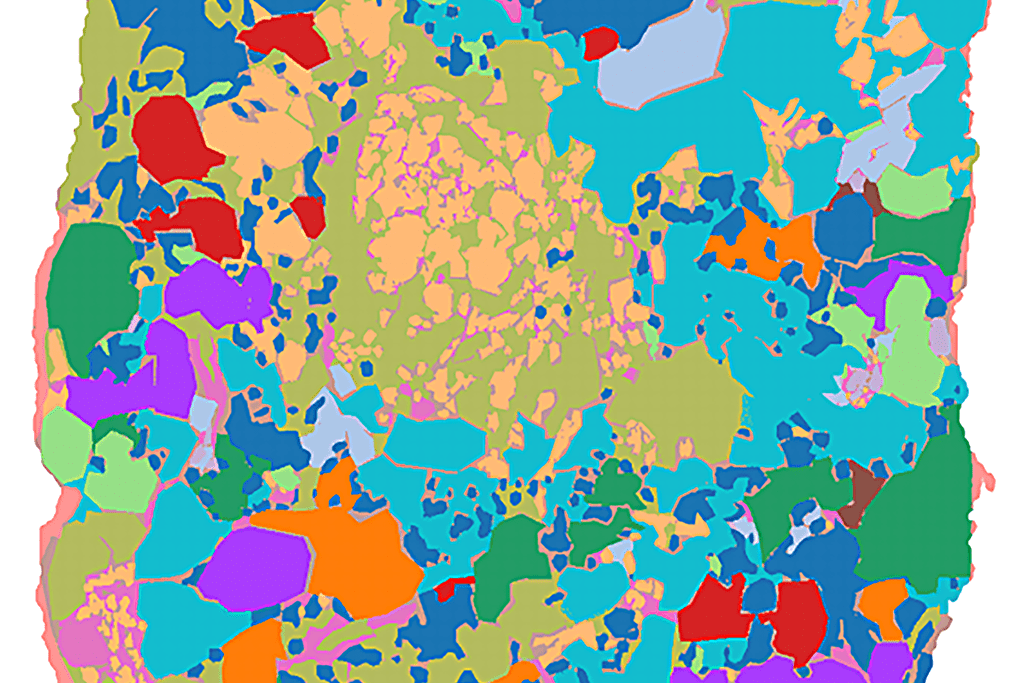
Photo: From the study