
What makes a Norway spruce shoot become a cone?
SciLifeLab researchers Stefania Giacomello and Sami Saarenpää, together with their colleagues Jens Sundström and Nathan Zivi at the Swedish University of Agricultural Sciences (SLU), have mapped the molecular changes behind Norway spruce reproductive development in a recent study published in Cell.
A Norway spruce (Picea abies) shoot starts out looking much like any other. As it develops, it can take one of two very different paths: it can become a vegetative branch or develop into a cone.
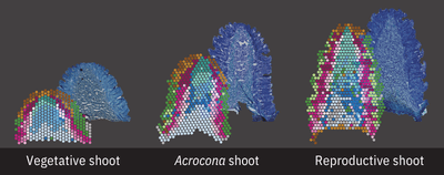
Researchers have now followed that process at the molecular level, using spatial transcriptomics to map gene activity across 88 shoot tissue sections from wild-type Norway spruce and its naturally occurring acrocona mutant. The samples covered three stages of development, allowing the team to see how initially similar structures gradually acquired either vegetative or reproductive identities.
The study identifies genes and regulatory networks associated with seed cone formation, including a previously uncharacterized MADS-box gene, DAL55. The researchers also propose a regulatory model connecting several transcription factors involved in reproductive development.
For Stefania, the project started with a long-standing fascination with a species whose biology still holds plenty of unanswered questions.
“I find Norway spruce truly fascinating because so much of its biology is still a mystery. It belongs to an ancient lineage of seed plants, has a huge and complex genome, is an essential component of boreal ecosystems, and we still know little about the molecular mechanisms controlling its reproductive development.”
A natural experiment in the forest
One of the things that made Norway spruce particularly well suited to the study was its naturally occurring mutant called acrocona. The acrocona transition shoots can contain both vegetative and reproductive features, giving the researchers an unusual opportunity to study the two developmental paths within the same structure.
“The acrocona mutant, which has been found only in a few locations in Sweden, made this even more interesting because it produces structures with both vegetative and reproductive features.”
The team combined the spatial transcriptomic data with morphological information from the same tissues. That combination was essential for identifying regulators and understanding where they were active as the shoots developed.
It also made it possible to study the spatial expression patterns of YABBY genes, pointing to mechanisms controlling lateral organ polarity that may be evolutionarily conserved between gymnosperms and angiosperms.
“I find acrocona such a special and exciting mutant to work with. The transition shoots themselves are morphologically interesting, but the mutant trees also reach reproductive age in about 5 years compared to the wild type’s 25 years. I wish that the gene activity programs we discovered can be translated into applications for the forest industry and tree breeding,” says Sami Saarenpää, PhD student in Giacomello’s lab.
Zooming in on the biology
One of the most striking things for Stefania was the amount of structure that appeared in the data at different scales.
“One of the most fascinating aspects for me was seeing how much key conserved spatiotemporal molecular structure there is at the whole tissue section level and at the same time how much molecular diversity there is in tiny tissue portions like the lateral organs, also across time.”
She describes it as a matter of perspective. Broad, recurring patterns become visible when looking at an entire tissue section, while very different patterns emerge in smaller regions.
The acrocona shoots offered another particularly revealing view.
“It basically provides us with a natural experiment in which we could follow the transition between these two developmental identities within the same structure.”
Seeing that transition in both the morphology and the spatial gene-expression maps was one of the most striking parts of the study.
The researchers could also use unbiased gene co-expression analysis to identify candidate regulators by selecting specific structures to compare. This is an advantage of spatial transcriptomics: researchers can choose precisely which parts of a tissue to compare while retaining their spatial context.
Then came the part where the computational predictions could be tested experimentally.
“It was a very happy moment when we could experimentally characterize these computational observations and obtain results consistent with the regulatory model suggested by the spatial data.”
Sami Saarenpää adds: “For me one of the greatest moments was seeing the very specific gene expression pattern of this unknown gene that had come up in several of our analysis. It turned out to be a completely uncharacterized gene, DAL55. Even better, we were able to link this gene to other developmentally important MADS-box genes.”
A study with deep roots
The connection to Norway spruce goes back further than this particular study.
“A fun fact is that this study has very deep roots in my earlier work on the Norway spruce genome.”
While still in Italy, she studied how transposable elements shaped the spruce genome. That work connected her with researchers at SciLifeLab and the Umeå Plant Science Center involved in the Norway spruce genome assembly and eventually brought her to Sweden.
Her postdoctoral research then focused on developing Spatial Transcriptomics for plants. Norway spruce was among the first plant species the method was demonstrated on, and the work also began her collaboration with Jens Sundström at SLU.
“In a way, this study brings the story full circle: from exploring the complexity of the Norway spruce genome to using spatial technologies to understand the complex biology of Norway spruce.”
From location to function
The work provides a molecular and computational framework for studying conifer reproductive development and demonstrates how spatial transcriptomics can uncover developmental programmes in non-model organisms. It also provides the first spatiotemporal transcriptomic atlas of conifer reproductive organ development.
Now the researchers want to take the approach further.
“I’m truly fascinated by the paradigm ‘from location to function’ and how conserved it is between the animal and plant kingdom.”
One direction is to conduct perturbation studies on key genes identified within and outside the proposed regulatory model and examine their effects on downstream gene expression.
The other is to increase the spatial resolution. The current study was performed at 55-µm resolution, meaning individual measurements can contain multiple cells. New spatial technologies can have cellular or even subcellular resolution, potentially revealing previously unresolved cell populations and regulatory programmes involved in reproductive development.
“Cellular resolution is an exciting advancement for developmental biology. I would be particularly interested in identifying gradients of gene expression, especially in boundary regions of different tissues. This could reveal how different organs form and what type of transcriptional landscape is important for organ formation,” Saarenpää says.
Spatial transcriptomics has already provided a way into the biology of a plant with a large and complex genome. The researchers now want to see how far they can take it.
“I’m really eager to continue pushing the application of new spatial technologies to plants as most of them work primarily for animal samples, leaving out a huge component of our ecosystems.”