Accurate and cost-efficient use of AI for cancer detection
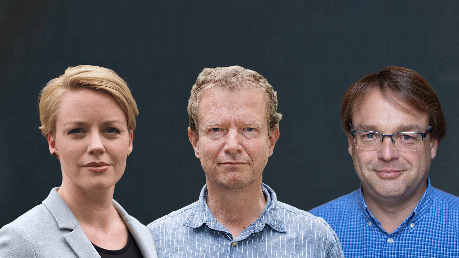
Researchers from SciLifeLab/KTH, Stanford University and Katholieke Universiteitte Leuven, have used a combination of advanced spatial transcriptomics and artificial intelligence (AI) to successfully detect cancer in histological images. Their results have been published in Nature Biotechnology.
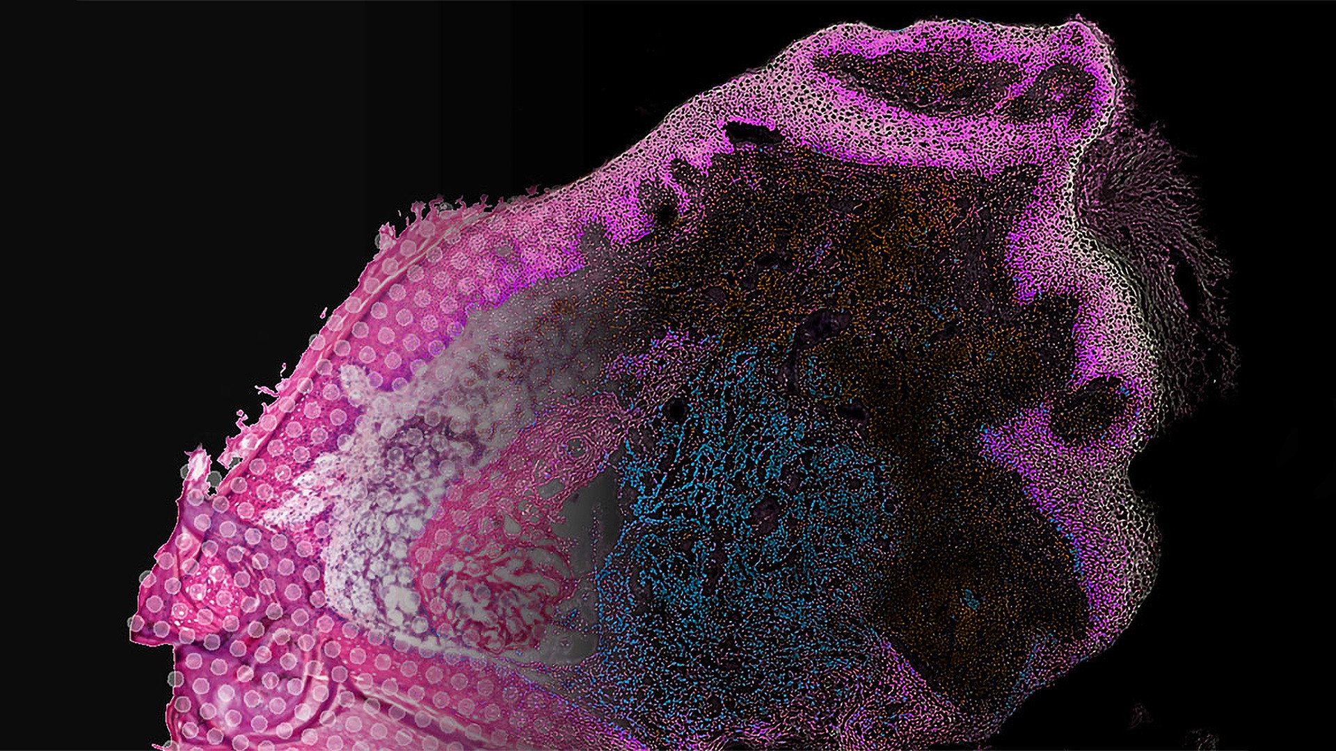
Artificial intelligence (AI) is an extremely useful tool, capable of distinguishing between healthy tissue and sick tissue by studying thousands of images from real life examples. At the same time computers become more powerful and the AI algorithms more complex, AI can diagnose patients just as fast, or faster, than doctors can.
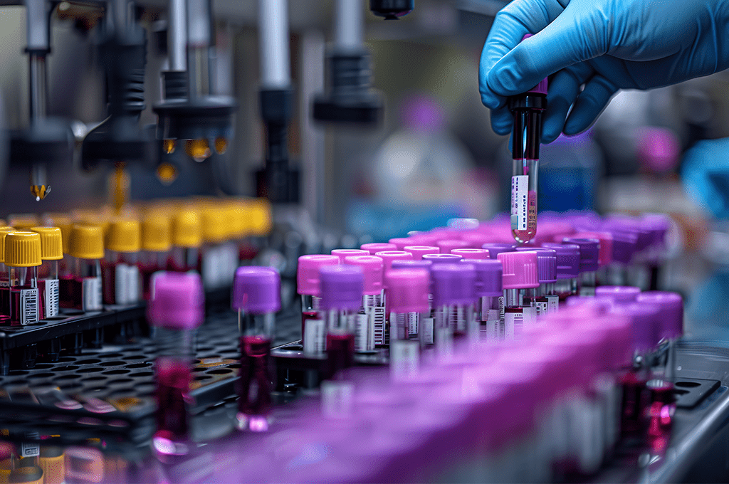
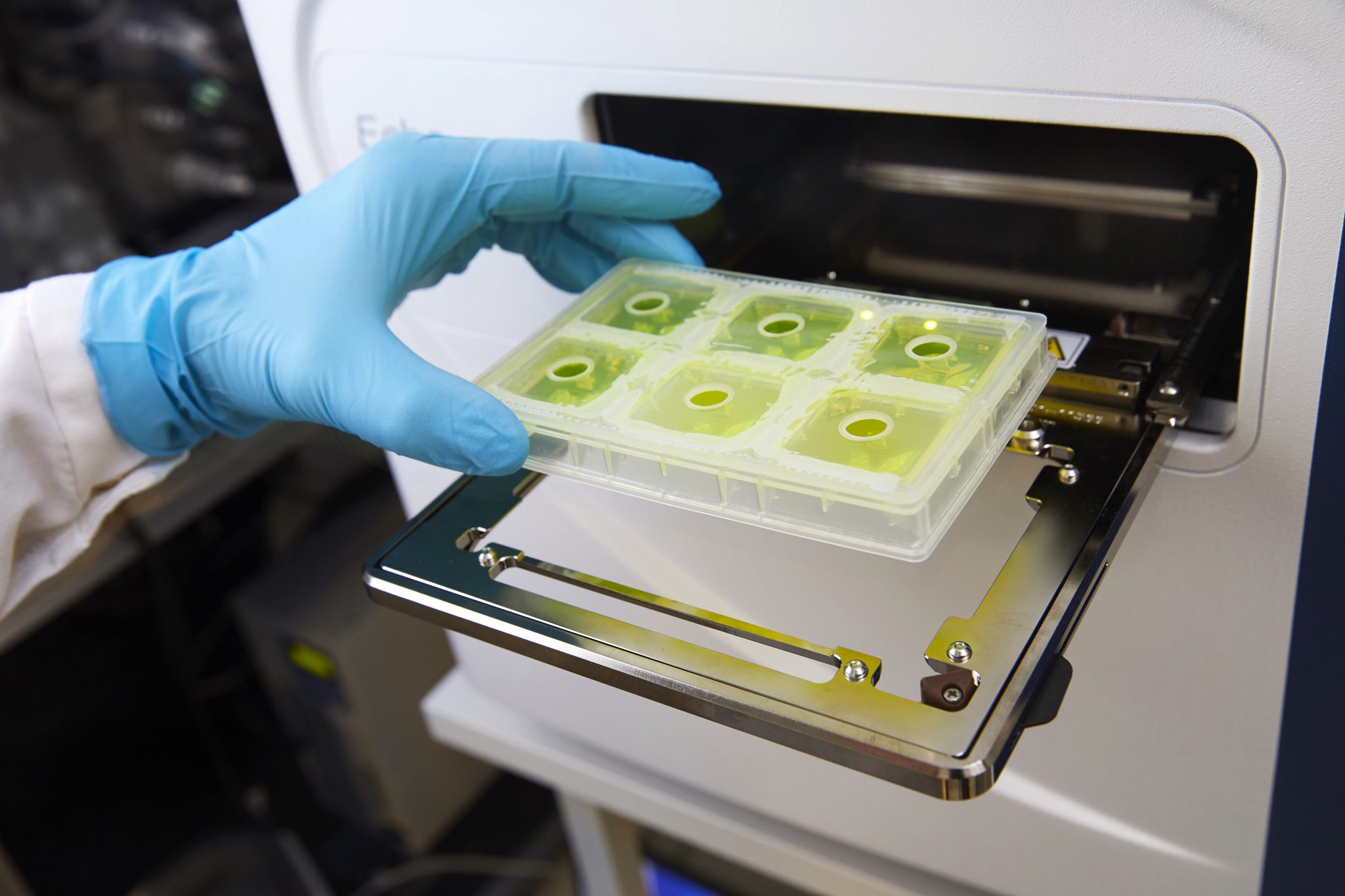
Now, researchers from SciLifeLab/KTH, Stanford University and Katholieke Universiteitte Leuven, have taken the technique even further by combining AI and advanced spatial transcriptomics, a technique capable of spatially visualizing the gene expression of cells in the tissue sample, providing the researcher with additional information about the location of specific cell types and how different cells can affect each other.
“Looking at tissue under a microscope has turned out to be successful and is today the most common way to diagnose several types of tumors. The limitation is that you can only look at the structure of cells”, says co-author Joakim Lundeberg (SciLifeLab/KTH), in a press release from KTH.
By combining histology images with gene expression data the researchers could provide the AI with a lot more information, making it more accurate in its diagnosis.
“By learning how histology is related to gene expression, we can improve the resolution of data from spatial transcriptomics”, says first author Ludvig Bergenstråhle (SciLifeLab/KTH).
The researchers have also developed a way to make the method both cheaper and faster by eliminating the expensive spatial transcriptomic step.
“The technique can be applied to histological images without associated gene expression data as long as there is enough data from reference experiments available. Because the histological images are less expensive to obtain than the gene expression data, the method makes it possible to analyze gene expression in much more tissue sections than previously possible. And knowing the gene expression is significantly more informative than just having histological images”, says Joakim Lundeberg.
He adds that the researchers can obtain the data just as quickly as the routine analysis of tissues in healthcare, but since they can also describe which genes are expressed and where in the samples, their analysis becomes much more accurate. In the long run, it can facilitate the next step, that is, what treatment to use but also improve early detection of tumors.
The research has been funded by the Cancer Foundation, the Knut and Alice Wallenberg Foundation, the Erling-Persson Family Foundation, the Foundation for Strategic Research, the Swedish Research Council and Leona M. and the Harry B. Helmsley Charitable Trust.