Mapping brain anatomy with groundbreaking molecular Artificial Intelligence
By utilizing AI and machine learning, researchers from SciLifeLab, KTH and Karolinska Institutet have developed a new way to create pictures of brain anatomy – more detailed than a microscope can provide. The results are published in the journal Science Advances.
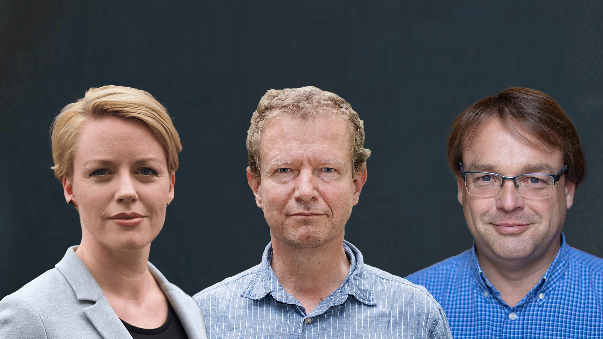
Up until now, visual differences in the organization of cells and nerves have primarily been used to create anatomical maps of the brain. These maps have played a huge role in the planning and execution of earlier research. But since they are created by different experts, not always using the same definitions, there has been some debate about their applicability, according to Konstantinos Meletis (Karolinska Institutet), lead author of the study now published in Science Advances.
In the study, which was conducted on the brain of an adult mouse, the researchers explored if there is a less subjective way to define and create brain maps using facts and data.
“To carry out this survey, we have used a method called spatial transcriptomics. It makes it possible to capture the molecules that encode the identity and function of cells”, explains Konstantinos Meletis, in a press release from Karolinska Institutet.
The new method for extracting RNA molecules has been developed by SciLifeLab researcher and head of the National Genomics Infrastructure, NGI, Joakim Lundeberg’s (KTH) group, in collaboration with Jonas Frisén at Karolinska Institutet. Currently, Joakim’s group is using the same method to study the human brain in cooperation with the Human Developmental Cell Atlas, HDCA.
The method is used to identify the exact position of RNA molecules in brain tissue.
Although the mouse brain is small, the large-scale survey with a total of 35,000 different measurement points, has been going on for almost three years. The mapping enabled the researchers to create a virtual 3D map of the entire mouse brain with information on over 15,000 genes, active in the various areas.
“What we have done is that we have replaced something that is very subjective – microscopy – with an objective molecular strategy”, says Joakim Lundeberg in a press release from KTH.
The method involves extracting, analyzing, and interpreting molecules of RNA, using AI and machine learning.
By capturing the molecular profile, without the need for knowledge of the brain or the function of the molecules, the researchers have been able to show that it is possible to recreate the entire brain’s detailed anatomy.
“When you look at a brain in a microscope you see different layers of the organ. With our method, you can see all genes that are active in each layer. But instead of, as with microscopy, having a subjective view of what is in an organ, we let the AI work out the pattern of an organ”, says Joakim Lundeberg in a press release from KI.
Read the press release from KI (in Swedish)
Read the press release from KTH (in Swedish)
Photo: Erik Flyg and KTH

