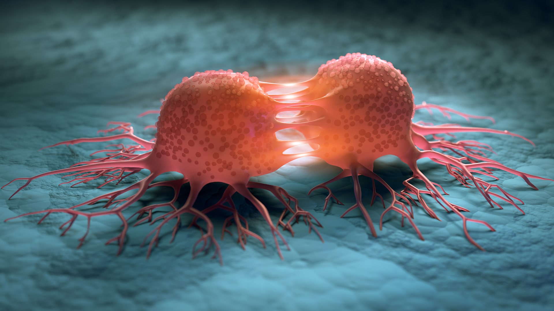
Mechanism behind cancer cell heat shock survival explained
In a recent study, a team of international scientists, identified some of the mechanisms used by human and mouse cancer cells to endure and survive heat shock. The results, published in Molecular Cell, revealed the mechanisms behind the phenomena but also how the cells are able to resume their original function afterwards, as well as passing the memory of stress along to their daughter cells.
“The results provide insight into the mechanisms that coordinate transcription in cells, which potentially could make a vital contribution in disease research”, says SciLifeLab Fellow and first author, Anniina Vihervaara (KTH), in a press release from KTH.
The researchers exposed embryonic fibroblasts and cancer cells to heat shock (42C) while monitoring their transcription activity across genes and their regulatory regions. When exposed to heat shock cells suffer from acute proteotoxic stress due to misfolding and aggregation of proteins. In an attempt to counter this, protein synthesis is reduced while the expression of chaperones, that can help existing proteins maintain their conformation, is increased. The heat shock response is involved in many diseases, such as cancer, Huntington’s disease and Alzheimer’s.
While embryonic mouse cells did not survive repeated or prolonged heat shock the cancer cells did. They even maintained their proliferation rate like nothing had happened.
“How cells survive heat shock is remarkable. The heat shock completely changed the transcriptional program of cells. Within minutes the cells switched to survival mode, inducing hundreds of genes, while repressing thousands more”, Vihervaara says.
The repressed genes are kept in a rapidly activatable state by pausing the transcription machinery at the early part of the gene. After the heat is removed, the cells recover and return to executing their cell-type-specific transcription program in a matter of hours, by simply turning on the transcription once again. Remarkably, the researcher could also observe how the cells transmitted the stress induced transcriptional memory to their daughter cells.
“Autophagocytosis-related genes were activated faster if the parental cells had experienced stress. These autophagocytosis pathway helps the cell to get rid of misfolded proteins and damaged organelles. Cancer cells also slowed RNA processing at the ends of the genes to reduce the burden for protein production”, explains Vihervaara.
A technique called Precision Run-On sequencing, which is able to closely monitor the progression of transcription at a nucleotide-resolution across the genome, was used in the study. This was followed by advanced data-analyses.
“Our aim is to bring this technical knowledge to physiological settings, where it can contribute to medical research. But first we need to understand the mechanisms of transcriptional reprogramming in model cell lines before we can understand them in physiological settings”, Vihervaara concludes.