Nanoscale 3D images of living mitochondria captured with novel technique
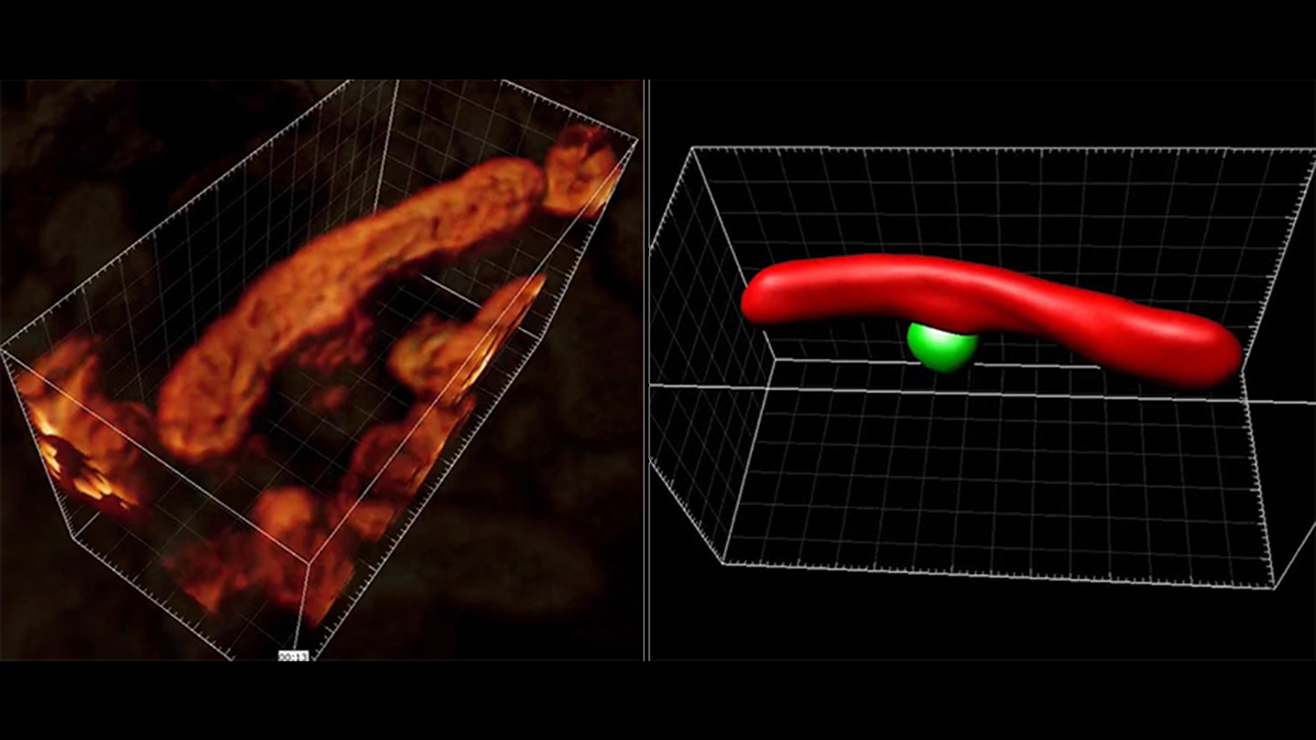
Researchers from SciLifeLab, KTH, and Karolinska Institutet have captured nanoscale 3D images of living mitochondria using a novel fluorescence microscopy technique. This is the first time researchers have been able to reconstruct entire living organelles from optical slices, capturing multiple images for reconstruction.
Until now, only electron microscopes have been able to create images with the same level of high resolution according to last author Illaria Testa, SciLifeLab fellow (KTH).
“This technique allows us to image proteins with a completely new level of 3D spatial details, and importantly in situ within the cell,” she says in an interview with KTH.
By optically “slicing” through an organelle, the researchers captured multiple images that could then be used to reconstruct it in 3D. In this way, they successfully generated a 3D image of living mitochondria, disentangling its mitochondrial network.
While you have to kill a cell when sectioning it for electron microscopy, the new technique named 3D pRESOLFT by the researchers delivers pictures on a wider scale and of living cells.
“Here, we don’t need to do that. The cells are happily moving and undergoing important functions,” Ilaria says. “Because the light is gentle we can look at living cells but with a new precision, that is, down to 50 nm (nanometres) in scale, or 20,000 times smaller than a human hair”.
“We now can see the 3D architecture of brain cells, and examine molecules that we consider important for learning and memory formation – and understand how they change location and shape when exposed to certain stimulations”.
The advance was reported in Nature Biotechnology and you can read the full press release from KTH here.