Optical super-resolution techniques shed light on neurons
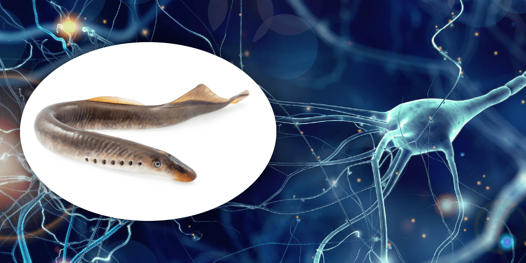
Using tissue expansion preparation and advanced microscopy, SciLifeLab fellow Ilaria Testa (KTH), in collaboration with Sten Grillner at the KI Department of Neuroscience, has shed light on spatial organization, abundance, and subcellular composition of neuronal subtypes in the lamprey spinal cord. Experiments performed in Ilaria Testa’s Advanced Optical BioImaging group, with support from the unit for Integrated Microscopy Technologies, reveal information about structure and function of neurons in tissue.
”The function of the two types of cerebrospinal fluid-contacting neurons that we have examined are of great importance to the field of neuroscience. Despite this, little has previously been known about their localization and function”, says first author Elham Jalalvand (KTH).
By applying tissue expansion microscopy combined with fast volumetric light-sheet imaging and super-resolution STED microscopy, the spatial organization, abundance, and subcellular composition of two distinct GABAergic CSF-c neuronal subtypes in the lamprey spinal cord were elucidated. One type – somatostatin – is pH sensitive and displays mechanosensitivity while the other – dopamine – only responds to mechanical stimuli. Furthermore, the two types of neurons use different cellular mechanisms for the transduction of mechanical stimuli to their cilia.
”Our imaging facility has supported with knowledge transfer in sample preparation to swell and make the tissue transparent and image this ‘super-separated tissue’ with fast volumetric light-sheet microscopy. says Steven Edwards, staff scientist at the Advanced Light Microscopy research infrastructure at SciLifeLab.
The study was partly funded by SciLifeLab SFO funding, and expertise and equipment at SciLifeLab’s infrastructure unit Integrated Microscopy Technologies was instrumental in facilitating the research effort.
The full publication, ExSTED microscopy reveals contrasting functions of dopamine and somatostatin CSF-c neurons along the lamprey central canal, can be found in eLife