Shedding light on mitochondria scaffolding proteins
Facilitated by SciLifeLab Advanced Light Microscopy unit (ALM), researchers from the Max Planck Institute for Biophysical Chemistry in Germany uncovered important information about the architecture of the mitochondrial inner membrane.
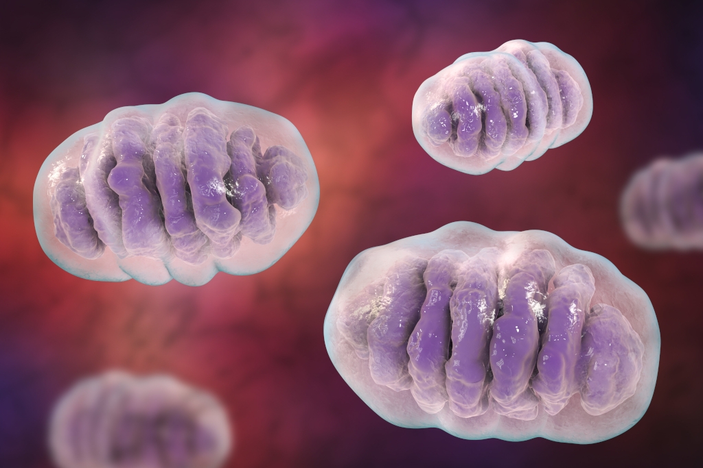
Mitochondria are double-membrane organelles working as cellular power plants and essential for all eukaryotic life. Their inner membranes are intricately folded so that the surface area towards the matrix, a gel-like substance within the inner membrane, is maximized. These folds are called cristae and are studded with proteins important for energy management. Recent discoveries have shown that the cristae are compartments rather than folds, connected to the transmembrane space by narrow channels called crista junctions.
The mitochondrial contact site and cristae organizing system (MICOS) is a complex that can be found at the crista junctions and plays a vital role for the establishment of a proper architecture of the mitochondrial inner membrane.
In a recent study, published in PNAS and facilitated by the SciLifeLab Advanced Light Microscopy unit (ALM), researchers demonstrated that Mic60, a subunit of the MICOS complex, as well as several of its interaction partners are arranged into intricate patterns in human and yeast mitochondria, suggesting an ordered distribution of the crista junctions.
The results opens the door to future microscopic investigations of the proteins responsible for scaffolding mitochondria.