A picture is worth a thousand words
On May 3, about 300 researchers participated during SciLifeLab Science Summit – Imaging the Complexity of Life. The day offered presentations, art exhibition and ample mingle opportunities.
This year’s SciLifeLab Science Summit focused on the impact of bioimaging, Whether on atomic or tissue level.
Movements within the cell
Both national and international speakers lined up to discuss the possibilities and future of imaging technologies.
Jennifer Lippincott-Schwartz, Howard Hughes Medical Institute Janelia, presented the fluorescence technology that her group uses to study cell architecture and dynamics. In particular, they color and track different organelles over time in living systems. For example the group tracks lipid droplets and their inter-organelle contacts over time, looking at their interaction with other labeled organelles.
By examining dynamics and volumes of organelles in 3D, the group can spot intimate relationships between e.g. mitochondria, endoplasmic reticulum (ER), lipid drops and other organelles.
“We segment out the mitochondria and examine which parts of the mitochondria is in contact with the ER. We can watch the movements over time, mapping out all the sites on the surface that the ER touches and hence, we believe, communicates with the mitochondria,” said Jennifer Lippincott-Schwartz.
The group has also investigated the dispersion of ER throughout the cytoplasm over time. Jennifer Lippincott-Schwartz showed several images of the ER and explained: “The ER occupies about 25 % of the cell excluding the nucleus at any particular time, but since it is moving, it explores 97 % of the cell within 15 minutes.
Art and science goes hand in hand.
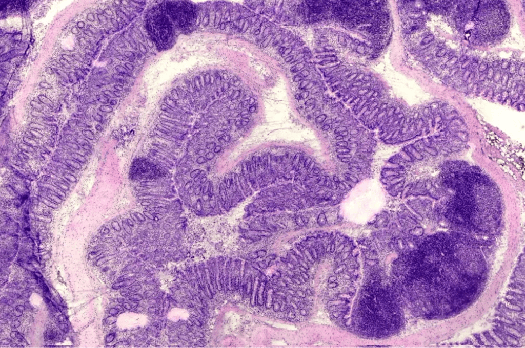
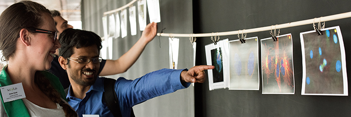
As per usual, SciLifeLab Science Summit featured an art exhibition. The Art Walk displayed beautiful microscope images from the SciLifeLab community. The art pieces captured chromosomes, cell division, plant hairs, close ups of insects, zebrafish eyes, and much more. A contribution by Matyas Molnar, displaying a light sheet microscopy image of a bug head, was voted the favorite by the conference participants.
Gaming for science
In his talk, Devin Sullivan, KTH Royal Institute of Technology, told the audience how it came to be that 200,000 gamers contributed to the Human Protein Atlas. He works with a group on the Cell Atlas, a high resolution subcellular atlas showing the spatial distribution of proteins within cells.
“We asked ourselves how we could analyze massive amounts of data with very little time and money. So, we turned to citizen science, which means that we allowed the general public to analyze scientific data.”
Devin Sullivan explained that the two biggest challenges with citizen science is to recruit the users and the cost to develop an engaging interface for them. To solve this the researchers teamed up with the game Eve Online, which have roughly 500,000 player.
In Eve Online the gamers analyzes mysterious ‘drifter DNA’ as a part of the bigger game. The players are presented with images and their job is to characterize protein locations in them. By performing these tasks the gamers gain awards in the game that can only be achieved by doing the analyses.
Several more researchers presented their work during the day, including protein synthesis at atomic resolution, bio-imaging opportunities at synchrotrons, multi-dimensional imaging during plant cell differentiation, how to use electron cryomicroscopy for in situ structural biology, and how structured illumination microscopy can offer insights into the regulation of mammalian meiosis.
Unit Corner
During the day, SciLifeLab imaging units presented their technologies and services at Unit Corner. In five condensed minutes they summarized the wide range of imaging technologies offered by SciLifeLab.
Highlight of the Year
Ten SciLifeLab publications from 2016 were nominated to Highlight of the Year. Each speaker presented their article in five minutes and the audience voted, judging both the scientific content and the presentation. The winner turned out to be the paper “Targeting SAMHD1 with the Vpx protein to improve cytarabine therapy for hematological malignancies” (DOI: 10.1038/nm.4265), presented by Sean Rudd.
Second place: ”Anoctamin 2 identified as an autoimmune target in multiple sclerosis” (DOI: 10.1073/pnas.1518553113), presented by Burcu Ayoglu
Third place: Accelerated cryo-EM structure determination with parallelisation using GPUs in RELION-2 (DOI: http://dx.doi.org/10.7554/eLife.18722), presented by Dari Kimanius.
The day ended with an informal mingle.