Key publications

Super-resolution stimulated emission depletion imaging of slit diaphragm proteins in optically cleared kidney tissue.
Kidney International 89(1), 243-247 (2016).
doi.org/10.1038/ki.2015.308

Confocal super-resolution imaging of the glomerular filtration barrier enabled by tissue expansion.
Kidney international 93(4), 1008-1013 (2018).
doi.org/10.1016/j.kint.2017.09.019

A molecular mechanism explaining albuminuria in kidney disease.
Nature Metabolism 2, 461-474 (2020).
doi.org/10.1038/s42255-020-0204-y

A Fast and Simple Clearing and Swelling Protocol for 3D In-Situ Imaging of the Kidney across Scales.
Kidney International 99(4), 1010-1020 (2021).
doi.org/10.1016/j.kint.2020.10.039

Three Dimensional Super Resolved Imaging of Paraffin Embedded Kidney Samples.
Kidney360 3(3), 446–454 (2022).
doi.org/10.34067/KID.0005882021

Deep learning–based segmentation and quantification of podocyte foot process morphology suggests differential patterns of foot process effacement across kidney pathologies.
Kidney International 103(6), 1120-1130(2023)
doi.org/10.1016/j.kint.2023.03.0

Hans Blom

Research Interest

My research interest concerns technical development and application of super-resolution microscopy-based imaging with a focus on applications in biology and medicine. Since 2014 the main focus has been to optically image kidney morphology across scales. Allowing for improved evaluation and understanding of pathophysiology in kidney filtration, which in the long run could enhance clinical renal applications and possible treatment strategies.

I am a docent in biological physics at the Royal Institute of Technology (KTH). As scientist I have acquired a unique competence in super-resolution fluorescence microscopy (postdoc in Nobel Laurate Stefan Hell’s lab, 2003-2005). I have nationally established an independent field of research in super-resolution microscopy since 2006 at KTH. My niche of applying super-resolution microscopy to life science has broadened the possibilities to visualize the nanoscale for collaborating scientists. As a super-resolution microscopy expert, I have during the last decade also built up the ALM unit, which provide access and training to state-of-the-art fluorescence imaging technologies at Science for Life laboratory in Solna. 

Imaging of kidney morphology across scales

In order to optically image kidney structures from the centimeter to nanometer scale we develop and apply optical clearing and swelling protocols combined with high-resolution fluorescence imaging. This allow us to in-situ image kidney structures in 3D across scales. See schematics and images below – also see key publications with technical explanations of the methods and its use to decipher early pathophysiological detection of kidney filtration structures.  


Left: Glomeruli and renal tubule structures [Podocin – green; E-cadherin – magenta] 
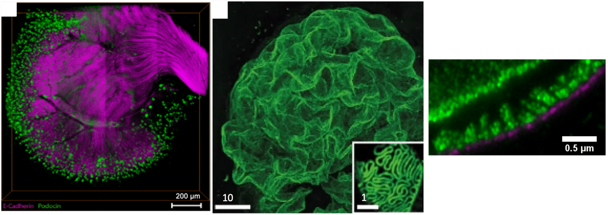
Middle: Glomerular filtration unit with podocyte foot processes [Podocin – green].
Right: Cross-view of glomerular filtration barrier [Podocin – green; Collagen IV – magenta]  


Project members & collaborators

  • David Unnersjö-Jess (KTH/University of Cologne) – Postdoc
  • Robin Ebbestad (KI/DS) – Clinical PhD-student
  • Jaakko Patrakka (KI) – National renal exper
  • Annika Östman Wernerson (KI) – National renal expert
  • Hannes Olauson (KI/KS) – National renal expert
  • Sigrid Lundberg (DS/KI ) – National renal expert
  • Thomas Benzig (NephroLab Cologne, Germany) – International renal expert

Contact

hblom@kth.se

Last updated: 2023-06-12

Content Responsible: David Gotthold(david.gotthold@scilifelab.se)