
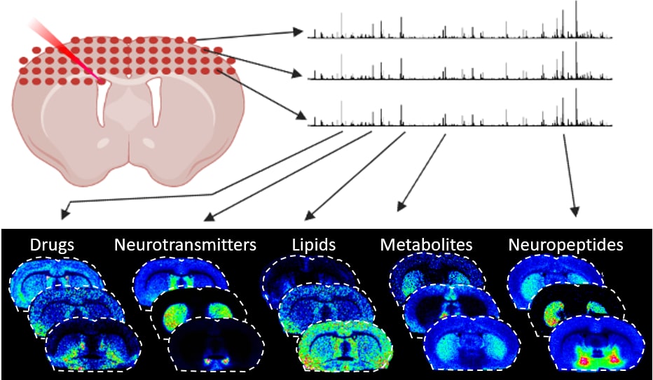
Spatial Mass Spectrometry
Spatial mass spectrometry is a label-free method that allows for the visualization and quantitative analysis of the distribution of targeted or untargeted chemical species directly in tissue sections. We utilize advanced mass spectrometry imaging (MSI) instruments and ionization methods, i.e., matrix-assisted laser desorption ionization (MALDI) and desorption electrospray ionization (DESI). The speed, sensitivity, and molecular specificity of the mass spectrometers enable the direct and simultaneous imaging of hundreds to thousands of molecules (in each pixel). The Spatial Mass Spectrometry unit provides a service for the imaging of drugs, their metabolites and endogenous biomolecules such as neurotransmitter systems, lipids, glycans, peptides, and small proteins directly in tissue sections at near-cellular lateral resolution.
Services
We perform everything from project design, sample preparation and imaging of tissue sections to optimize the project results by using bioinformatic tools. All projects are unique and might require individual method developments to different extents.
Proposed projects are assessed and prioritized according to the following model
Initial screen of project by unit
- Technical feasibility and method developments (stop/go decision)
- Capacity requested for the project (stop/go decision)
- Pilot study (stop/go decision)
Project prioritization is based on
- Scientific potential
- Significance of unit specific technique for project
- Supporting preliminary data from other techniques
- Supports unit development (competence and techniques)
- Feasibility of time lines
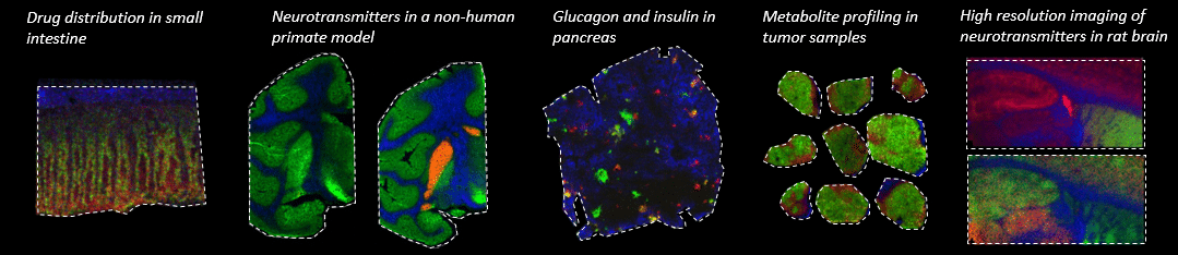
Applications
- ADME
MSI enables simultaneous and label-free quantitative analysis of drugs and metabolites across multiple tissue types.
- Safety toxicology
MSI provides an option for tissue classification at the molecular level and could thus be very useful as an alternative or complement to histology for the diagnosis and characterization of toxicological findings.
- Drug Blood-Brain Barrier (BBB) Permeability
MSI visualizes and quantifies the brain distribution of drugs with varying blood-brain barrier permeability, determines the blood-brain barrier permeability and defines the drug distribution in very small brain structures.
- Neuroscience
MSI provides a strategy for simultaneously imaging, identifying and quantitating the absolute concentrations of multiple neurotransmitters and neurotransmitter precursors and metabolites directly in brain tissue sections.
- Oncology and Pathology
MSI has the ability to detect and characterize tumour cells and their environment in a spatial context and can be a complement to aid pathologists and clinicians.
Equipment
Mass spectrometry instruments
- SolariX 7T-2ω Fourier-Transform Ion Cyclotron Resonance (FTICR), Bruker Daltonics
The two solariX MALDI instruments allow for the mapping of the distribution of drugs and metabolites, neurotransmitters, neuropeptides and lipids. The FTICR instruments offer ultrahigh mass resolving power that maximizes the number of analytes detected. They require tissue sections to be placed on conductive glass slides.
- timsTOF flex MALDI-2, Bruker Daltonics
The timsTOF flex instrument provides enhanced sensitivity, featured by a two-laser MALDI-2 technology, increased speed, high lateral resolution (5 µm), and trapped ion mobility separation capabilities and can be used for a broad variety of MALDI-MSI applications. Requires tissue sections to be placed on conductive glass slides.
- MALDI/DESI Q-Tof Synapt G2Si, Waters Corp.
The MALDI SYNAPT G2-Si is suited for analysis of tissue sections placed on regular microscope glass slides (non-conductive). Lateral resolution of 20 µm.
- DESI Xevo TQ-XS Triple Quadrupole Mass Spectrometry, Waters Corp.
Analysis with the DESI Xevo TQ-XS instrument allows targeted imaging of small molecules on regular microscope glass slides (non-conductive) at a lateral resolution of 20 µm.
Matrix application and tissue cutting instruments
- TM-Sprayer and M3+ Sprayer (HTX-Technologies) for MALDI matrix application
- MALDI matrix sublimation system
- CHIP-1000 Chemical Printer, Shimadzu Corp. for application of standard curves
- BioSpot Benchtop workstation, BT 600, BioFluidix for application of standard curves
- Cryostats, Leica CM1900 and CM3050, Leica Microsystems
Data analysis and bioinformatics tools
- msIQuant (developed in-house)
- SCiLS lab
- Metabolite identifier (developed in-house)
- Statistical analysis (GraphPad Prism and SIMCA)
- FlexImaging
Using the unit
Please send us an email with questions, suggestions or project proposals using the contact emails provided. We will get back to you shortly after to schedule a meeting for further discussions.