
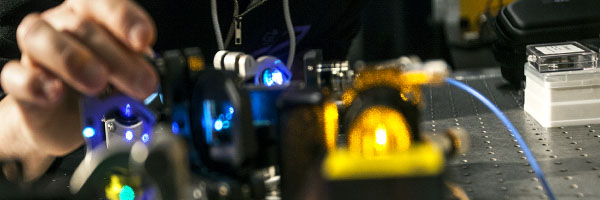
Integrated Microscopy Technologies (IMT)
Follow links and information below to access technologies supported through the Integrated Microscopy Technologies (IMT) infrastructure unit, which include Correlative Array Tomography (CAT), Nanoscale Secondary Ion Mass Spectrometry (NanoSIMS), Focused Ion Beam Scanning Electron Microscopy (FIB-SEM) and Advanced Light Microscopy (ALM).
–> APPLY FOR IMT SUPPORT here LINK.
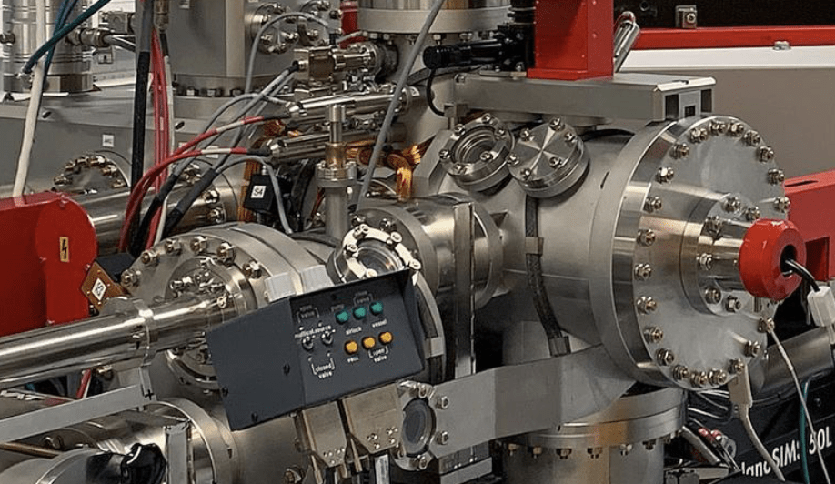
IMT-Gothenburg: CAT & NanoSIMS
Correlative array tomography (CAT) combines microscopy modalities to explore the 3D cellular architecture of large samples in extremely fine structural and molecular detail by, for example, merging the molecular discrimination of light microscopy with the ultrastructural imaging of electron microscopy.
LINK to CAT services at University of Gothenburg.
Nanoscale secondary ion mass spectrometry (nanoSIMS) offers a secondary ion nanoprobe to carry out nanoscale projects, a unique technology in the Nordic region, to address questions in materials, geological, marine, environmental, medical and life science fields.
LINK to NanoSIMS services at University of Gothenburg.
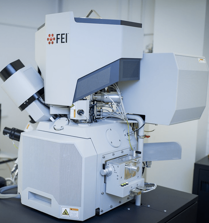
IMT-Umeå: FIB-SEM
Focused ion beam scanning electron microscopy (FIB-SEM) reveals subsurface structural detail by making precise cuts with a FIB, then imaging the exposed surface with a high-resolution SEM.
LINK to FIB-SEM services at Umeå University.
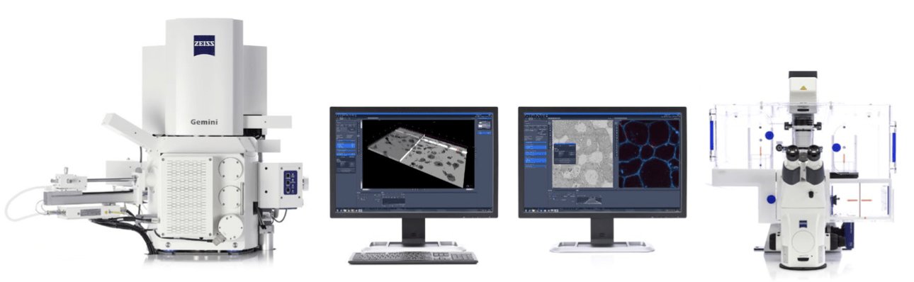
IMT-Stockholm: Advanced Light Microscopy (ALM)
The mission of the IMT Stockholm unit / Advanced Light Microscopy (ALM) facility is to give support with nanoscale biological visualisation. In addition we support molecular analysis by fluorescence correlation spectroscopy methods and superresolution additions for nanoscale dynamics. Moreover light-sheet fluorescence microscopy allow users to image live and/or optically cleared larger samples at unprecedented volumetric speed with low phototoxity. Single cell ultra-fast volumetric imaging of biological processes at high-resolution is further provided by live lattice light-sheet microscopy.
Apply for ALM support via our national project portal LINK.
Services (ALM)
Support is done to all stages of the project which can include (pre)planning, sample optimisation, fluorescent probe selection, image acquisition, and initial post-acquisition image processing – focusing on:
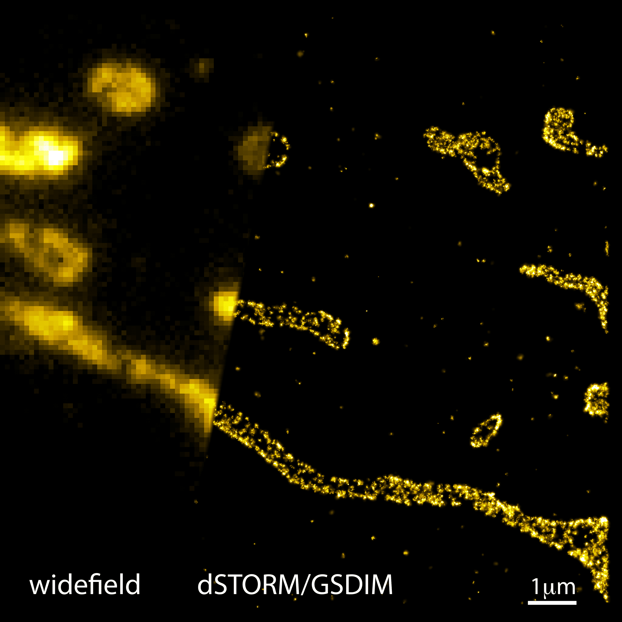
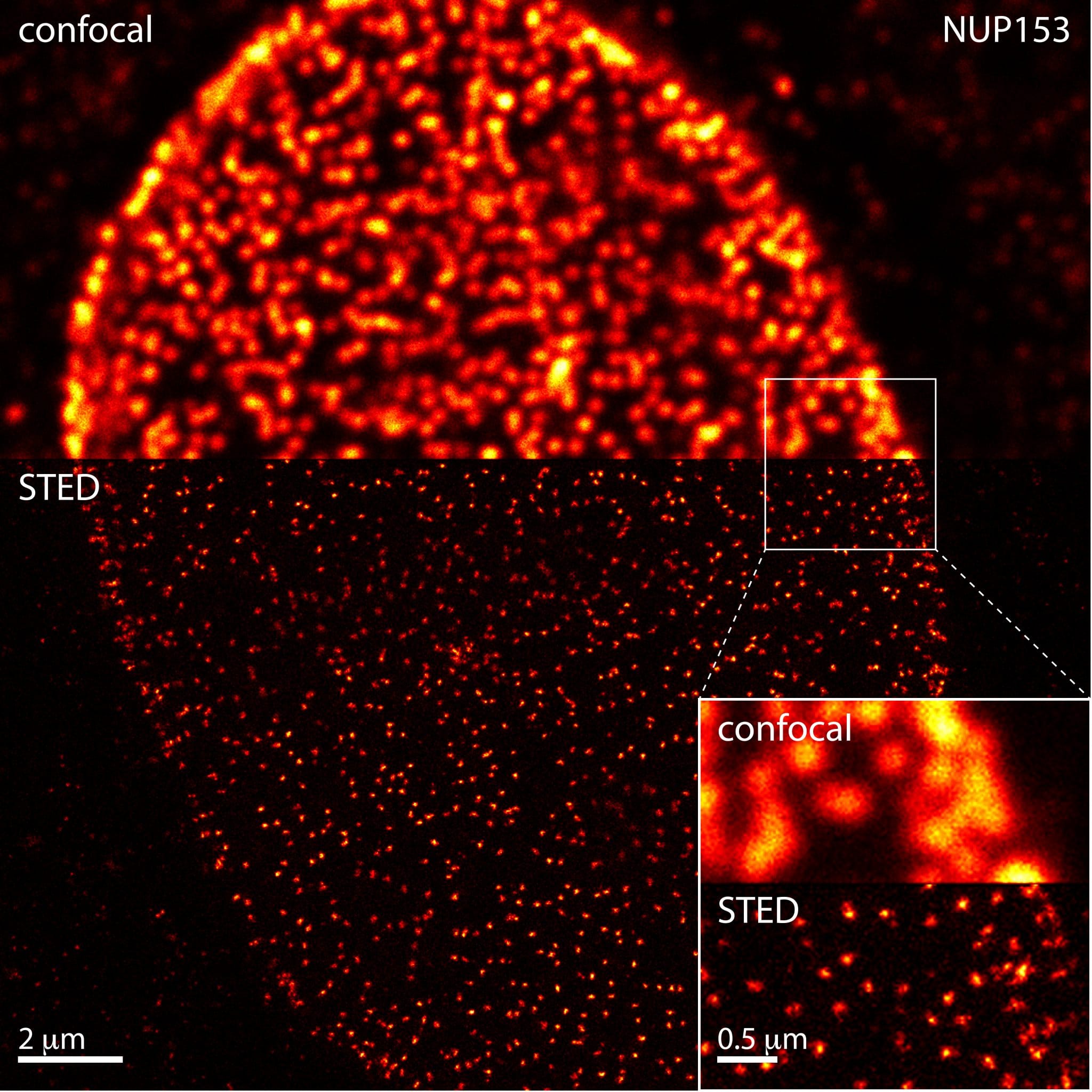
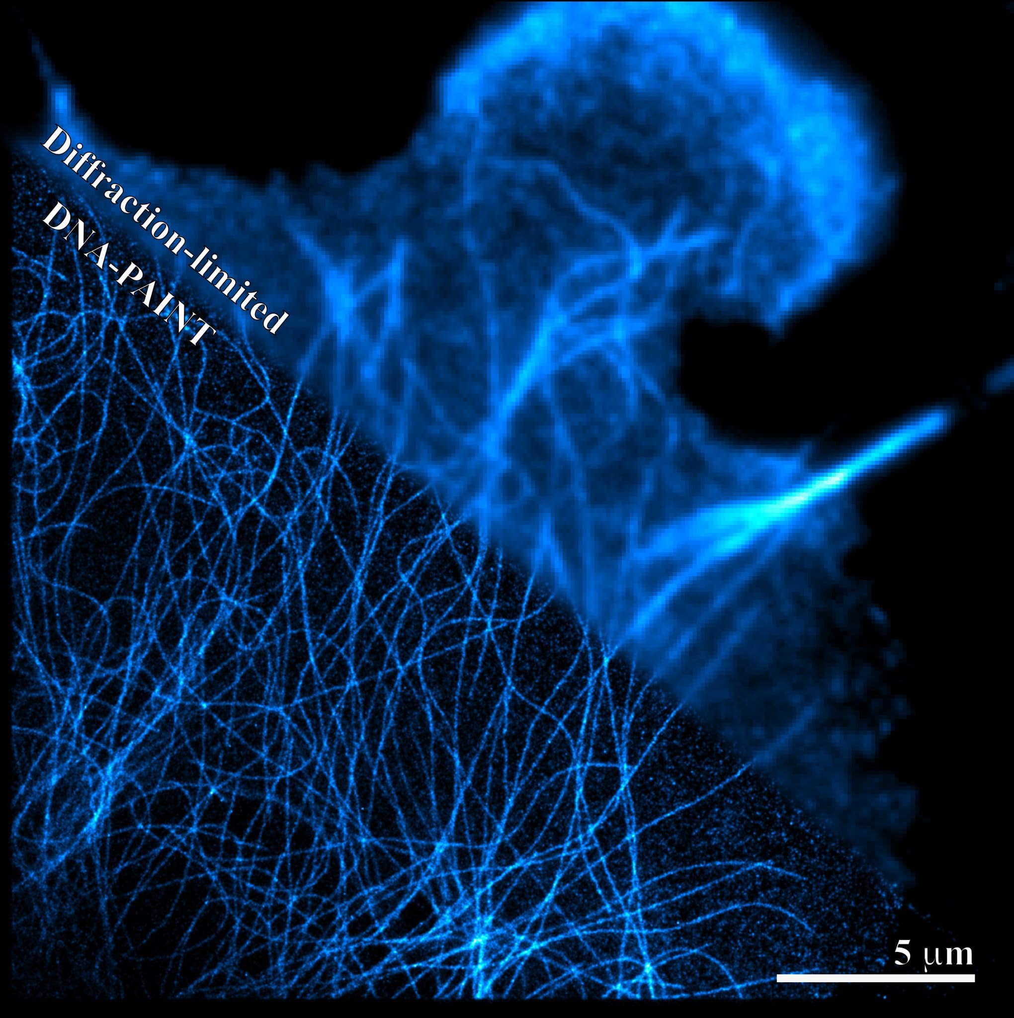
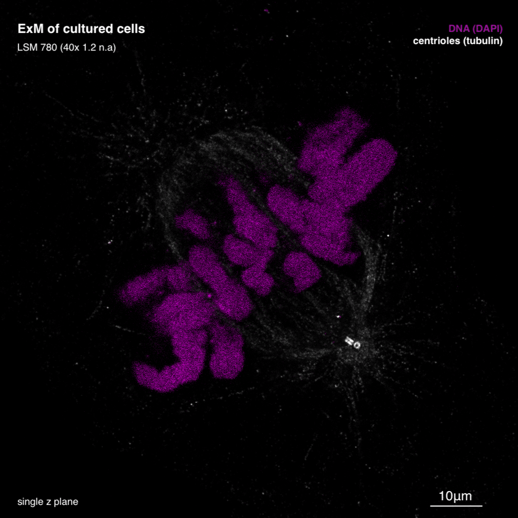
- Super-resolution microscopy // nanoscale imaging of fixed cells incl. cleared or expanded tissue and cells.
- Fluorescence Correlation Spectroscopy // single molecule dynamics in-vitro or in-situ.
- MINFLUX tracking // ultra-fast nanoscale dynamics of proteins and lipids in cells.
- Light-sheet microscopy // volumetric imaging of organoids, model organisms and cleared tissues.
- Lattice light-sheet microscopy // fast high-resolution volumetric imaging of cellular processes.
Transfer of unique knowledge to individual researchers are supported nationally, including organization of workshops and courses in advanced fluorescence imaging and spectroscopy applications and method developments.
IMT Stockholm (ALM) overview on support: https://youtu.be/mqx49azmqtY
Methods (ALM)
SIM/STED/SMLM/ExM/MINFLUX imaging
- Investigation of molecular architecture of sub-cellular entities.
- Multicolour molecular organisation with sub-diffraction resolution imaging.
FCS/FCCS/FRET-FCS/Dynamic Profiler/STED-FCS/MINFLUX tracking
- Analysis of protein-protein, protein-peptide/ligand and protein-vesicle interactions
- Aggregation and affinity analysis at single molecule concentration levels
- Molecular nanoscale tracking and dynamic analysis in living cells
Light-sheet/Clearing/Lattice Light-sheet
- Live: Embryogenesis and Organogenesis (e.g. in Zebrafish)
- Live: 3D cell culture imaging using spheroids, tissue and organotypic culture
- Imaging optically cleared/expanded samples (e.g. CLARITY, CUBIC-R/X, ExM)
- Rapid, sub-cellular volumetric imaging of biological processes (LLS)
Equipment (ALM)
- SMLM. Zeiss ElyraPS1/7 3D PRILM
- STED. Leica SP8 3X STED with FALCON FLIM/FCS
- SIM/SIM2. Zeiss ElyraPS1/7 3D SIM
- AIRY2/FCS. Zeiss LSM980
- FCS. Zeiss LSM780
- Light-sheet. Zeiss Z.1/7
- Lattice light-sheet. Zeiss LL7
- MINFLUX. Abberior Instruments 2D/3D dual-color
Get support from the ALM unit
Apply for support via our national project portal LINK.
International users may also apply through the Euro-BioImaging project portal.
Image gallery (ALM)
Video gallery (ALM)
Zebrafish embryo development acquired on the Zeiss Light-Sheet Z.1
Mailing Address (IMT Stockholm)
SciLifeLab
ALM, Gamma 3
Box 1031
171 21 Solna
Sweden
Visiting Address/Deliveries (IMT Stockholm)
ALM/Scilifelab gamma-3
Tomtebodavägen 23A/23B
171 65 Solna
Sweden