Data Driven Systems Biology: Harnessing Big Data and Systems Approaches to Decode Complex Biology
March 24, 2026 @ 09:00 – March 25, 2026 @ 17:00 CET
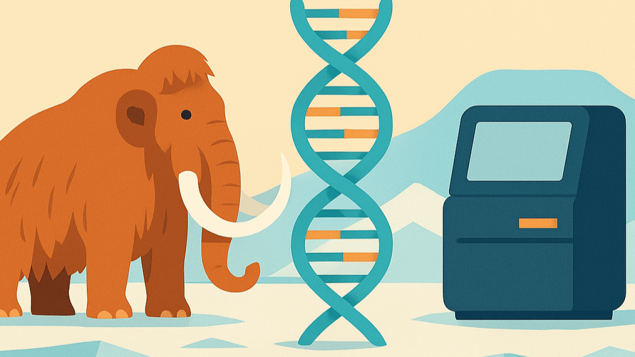
In the era of high-throughput technologies and rapidly expanding biomedical datasets, the field of systems biology is undergoing a transformative shift. The Data-Driven Systems Biology conference brings together leading researchers who are leveraging computational, statistical, and systems-level approaches to integrate and interpret complex biological data. This conference will explore how multi-omics, single-cell technologies, and spatial profiling, combined with advanced computational modeling and machine learning, are reshaping our understanding of dynamic biological systems.
The DDLS research area symposia series aims to engage and build a strong national scientific community around the DDLS research themes. Each of the four areas arranges two symposia per year. Everyone interested in data-driven research is welcome to take part. We aim to unite researchers, industry, and healthcare to foster collaboration and advance the frontiers of data-driven life science.
Target Group: The DDLS research area Expert Group in Cell and Molecular Biology invites all interested in Data-driven life science to meet, present, interact, and discuss Imaging in Cell and Molecular Biology.
The event will take place at Life City, Solna, Stockholm, and will include presentations from international and national invited speakers and selected abstracts. The event is free of charge.
Date: March 24-25, 2026
Start on March 24: 11:00 – 12:30 Registration open. 11:30-12:30 Network lunch. The conference starts in the Lecture hall at 12:30.
End: March 25 with a Network lunch from 12:30 to 13:30.
Venue: Life City, Solnavägen 3H i Solna.
Organized by: Arne Elofsson and Eduardo Villablanca, DDLS Expert Group in Cell and Molecular Biology.
Contact: events@SciLifeLab.se
Program
Poster session
The poster session will take place at 17:30 in Delta, Campus Solna. Light food and drinks will be served. Please hang your poster on any empty poster board as soon as you arrive to Campus Solna after the Conference’s first day. Pins will be available.
Registration
The registration deadline is March 10. We cannot accept any posters after deadline. To avoid empty seats, registration will remain open with a que-list until the event begins. However, registering after March 10 requires you to write your name on a name tag at on-site registration. Unfortunately, we cannot accommodate allergies or dietary preferences for those who register after March 10.
Cancellation
Please! To minimize empty seats and food waste, cancel your registration if you are unable to attend, or update your lunch selection if your attendance changes. Cancel/update via the Confirmation email or email events@scilifelab.se.
Confirmed speakers
Title: Data, Digital Twins and AI
Bio: Prof. Alfonso Valencia is ICREA research Professor, Director of the Life Sciences Department of the Barcelona Supercomputing Center, Director of the Spanish National Bioinformatics Institute INB/ELIXIR-ES and coordinator of the data pillar of the Spanish Personalised Medicine intiative, IMPaCT. His research interest is the development of Computational Biology methods and their application to biomedical problems. Some of the computational methods he developed are considered pioneering work in areas such as biological text mining, protein coevolution, disease networks and more recently modelling cellular systems (digital twins). He participates in some of the key cancer related international consortia. In terms of community services, he is one of the initial promoters of the ELIXIR infrastructure, founder of the Spanish and International Bioinformatics networks and former president of ISCB, the international professional association of Bioinformaticians. He is Executive Editor of the main journal in the field (Bioinformatics OUP).
Abstract: In this talk I will treat in some order these three topics: Data, human Ditigal Twins and the impact of AI in biomedicine.
I will address the persistent bottleneck of data access in biomedical research, where the combination of legal and technical hurdles span the entire data lifecycle, from discovery and access to integrated analysis. I will mention the current developments to overcome these limitations by implementing federated discovery and analysis systems designed to work across borders and heterogeneous resources.
Regarding Digital Twins, I will discuss the importance of those developments in the transition from statistical correlations, which are standard in genomics analysis, to mechanistic interpretations that will better align with the core objectives of molecular biology. We are approaching this underdeveloped area with the construction of mechanistic models of cellular systems, that are already showing promising results in critical biological systems.
Finally, I will discuss how the rapid advances of AI is influencing the work in different areas of biomedicine – including specific examples of how we combine Digital Twins and AI methods- as well as what I see as promises and limitations in this area.
Title: From Omics to Mechanisms: Deep Learning Models of Molecular Networks for Precision Cancer Medicine
Bio: Avlant Nilsson is an Assistant Professor in Precision Medicine at the Department of Cell and Molecular Biology, Karolinska Institutet, and a group leader at SciLifeLab through the DDLS program. He holds an MSc (2014) and a PhD (2019) in Biological Engineering from Chalmers University of Technology, where his thesis focused on the metabolism of proliferating cells, including liver cancer. He then pursued postdoctoral research at the Massachusetts Institute of Technology (2019–2023), developing neural network models of signal transduction in immune cells. His research group, currently comprising of two PhD students and two postdoctoral researchers, develops data-driven models of molecular networks to understand how genetic alterations, cell type of origin, and cell–cell interactions shape cancer biology. The long-term goal of the lab is to advance computer-aided design of cancer medicine by predicting drug responses, resistance mechanisms, and microenvironmental interactions.
Abstract: Cancer is highly heterogeneous, spanning a multitude of genetic alterations, cell types, and microenvironmental contexts, making it difficult to identify effective treatments for individual patients. Deep learning models are powerful predictive tools that could be applied to large-scale molecular data, but their black-box nature limits their ability to generate mechanistic insight to guide therapeutic intervention.
To overcome this, we develop biologically informed neural network models that embed known molecular interaction networks directly into the deep learning architecture. Specifically, we construct recurrent neural network models of cells in which biomolecules are represented as nodes with connections defined by their physical interactions. These models take data with molecular causes as input (such as mutations and copy number variations) and are trained to predict omics readouts, including gene expression, protein phosphorylation states, and metabolite levels.
By training models on high-throughput datasets spanning different cell types, perturbations, and conditions, we can predict molecular responses in conditions that are withheld during training. We also use these models to expose non-canonical signaling events that would be difficult to identify directly from the data using standard analysis approaches. With this, our framework offers the potential to identify novel drug targets, biomarkers, and to predict resistance mechanisms.
Title: Spatially resolving B cell clonal dynamics in cancer and beyond
Bio: Dr. Camilla Engblom is a SciLifeLab Fellow and an Assistant Professor in the Division of Immunology and Respiratory Medicine and the Department of Medicine, Solna at the Karolinska Institutet (KI). Dr. Engblom received her PhD in Immunology from Harvard University in 2017 focusing on long-range cancer-host interactions involving myeloid cells (Dr. Mikael Pittet’s lab at Massachusetts General Hospital/Harvard Medical School). As a MSCA postdoctoral fellow in Dr. Jonas Frisén’s lab (KI), Dr. Engblom developed a spatial transcriptomics-based tool (Spatial VDJ) to map B cell and T cell receptors within human tissues. Located at SciLifeLab and the Center for Molecular Medicine (KI), the Engblom lab’s main research focus is to spatially and functionally resolve B cell clonal dynamics in cancer tissues and beyond.
Abstract: B cells perform functions critical to human health, including antibody production and antigen presentation. B cells develop, differentiate, and expand in spatially distinct sites across the body. B cells express clonal heritable B cell receptors (BCR) that confer exquisite molecular (i.e., antigen) specificity. B cell receptors can be defined by sequencing. Linking specific BCR sequences to their molecular and cellular surroundings, i.e., ‘clonal niche’, could help us understand and harness B cell activity. A technological bottleneck has been to capture the location of BCR sequences, and by extension B cell clonal responses, directly within tissues. We recently developed a spatial transcriptomics-based approach (Spatial VDJ) and associated computational pipelines to reconstruct B cell clonality in human tissues. Here, we present adaptation of Spatial VDJ to murine tissue to enable preclinical studies and B cell receptor dynamics under inflammatory conditions, including cancer.
Title: Enabling technologies for spatial metabolomics: Moving from single cells to 3D-space exploration in mixed reality
Bio: Carsten Hopf obtained his PhD in biochemistry from Tübingen University/Max-Planck-Institute for Developmental Biology. As an EMBO fellow in neuroscience, he then worked at the Johns Hopkins University School of Medicine for three years, before joining Cellzome AG, a proteomics-focused drug discovery platform company in Heidelberg in 2001. There, for 13 years, he served in multiple roles in platform technology, assay development, drug discovery and business development, and eventually as part of Cellzome’s leadership team until the end of 2014.
Since 2005, Carsten Hopf is a professor of bioanalytics, proteomics and drug discovery at TH Mannheim. He currently heads TH.M’s CeMOS Research and Transfer Center that was recently selected as mass spectrometry imaging partner site of the EU-OPENSCREEN research infrastructure that SciLifeLab and KI are also parts of. Carsten is the Speaker of the M2Aind partnership for innovation in health industry in Mannheim, and he serves on various science and innovation cluster boards. He is also an associated professor in the Medical and Biosciences Faculties of Heidelberg University and co-chair for “imaging” of the German Society for mass spectrometry (DGMS). Carsten’s research focuses on Mass Spectrometry and Optical Spectroscopy enabling technologies for health and life science, especially spatial systems biomedicine.
Abstract: MALDI-Mass spectrometry imaging (MSI), also referred to as spatial metabolomics, has emerged as a powerful technology for spatially resolved analysis and visualization of lipids and metabolites in systems biology and clinical research. Advancement of MSI requires rapid progress in multiple areas such as instrumentation, experimental workflows and computational strategies to harness big data. The talk will therefore initially review a “classic” technology show case using tissue slices: Spatial metabolomics revealed that Tet3 knockout enterocytes exhibit an unphysiological metabolic profile when compared with their wild-type counterparts suggesting that terminal cell differentiation is regulated by TET3 at the metabolic level. MSI technology has recently moved into two new directions: Single-cell metabolomics and 3D-reconstructed metabolomics.
To study proinflammatory activation of iPSC-derived microglia by bacterial lipopolysaccharide (LPS), we developed the PRISM-MS (PRescan Imaging for Small Molecule – Mass Spectrometry) platform for analysis and on-cell MS2 identification of low mass metabolites (<200 Da) in large cell populations. Itaconate and taurine were identified as markers for “activated” versus “resting” microglia, respectively. Translation of single cell results to endogenous microglia in organotypic rat hippocampal slice cultures indicated that LPS-activation involves changes of the itaconate-to-taurine ratio and alterations in neuron-to-glia glutamine-glutamate shuttling.
To investigate fibroblast-colon cancer cell interactions in a simple 3D-culture model and in patient-derived organoids (PDOs), we built a translational 3D MSI platform as an end-to-end solution for 3D-enabling sample preparation, 3D-reconstruction and data processing, 3D-rendering, and immersive user interaction with organoid big data in a mixed reality. When applied to colon cancer PDOs, the methodology revealed that fluid-filled cysts characteristic of these PDOs were rich in purine nucleotides.
Title: Harnessing Big Data for Network Biology with FunCoup 6
Bio: Erik Sonnhammer is Professor of Bioinformatics at Stockholm University, and previously had the same position at Karolinska Institutet, Stockholm. He did a Ph.D. in bioinformatics at the Sanger Institute in Cambridge, England. His research interests are in network and systems biology to understand gene and protein function on a large scale. The group has made many contributions to Gene Regulatory Network analysis, including inference, benchmarking, and simulation.
Abstract: FunCoup 6 is a major update to the FunCoup network database, providing researchers with a significantly improved and redesigned platform for exploring the functional coupling interactome. The FunCoup network database (https://FunCoup.org) contains some of the most comprehensive functional association networks of genes/proteins available. Functional associations are inferred by integrating different types of evidence combined with orthology transfer. FunCoup’s high coverage comes from using ten different types of evidence, and extensive transfer of information between species.
Key innovations in release 6:
– Enhanced regulatory link coverage: FunCoup 6 now includes over half a million directed gene regulatory links in the human network alone. 13 species in FunCoup now contain regulatory links..
– The website is completely redesigned, with updated API functionalities, enhancing user accessibility and experience.
– Integrated advanced online tools for network analysis: The integration of TOPAS for disease and drug target module identification, along with network-based pathway enrichment analysis using ANUBIX, expand the utility of FunCoup 6 for biomedical research.
– New training framework: applied to produce comprehensive networks for 23 primary species and 618 additional orthology-transferred species.
– FunCoup 6 is also available as a Cytoscape app.
A unique feature of both the FunCoup website and the Cytoscape app is the possibility to perform ‘comparative interactomics’ such that subnetworks of different species are aligned using orthologs. FunCoup further demonstrates superior performance compared to other functional association networks, offering researchers enhanced capabilities for studying gene regulation, protein interactions, and disease-related pathways.
Title: Learning cellular dynamics of tissues from single-cell and spatial omics
Bio: Jean researches mathematical rules in the molecular tricks that cancer cells use to escape destruction by immune cells. We seek to articulate the molecular chat between immune and cancer cells into equations, to serve as the foundation to engineer personalized cancer immunotherapy. We combine single-cell and spatial tumor profiling experiments, machine-learning & data science, and physics-style mathematical modeling.
Abstract: Cell proliferation and death rates are central to tissue biology but measuring them in vivo remains a persistent challenge. Here we present tissue dynamics inference (TIDYI), which quantifies absolute cell proliferation and death rates from non-longitudinal single-cell RNAseq snapshots. TIDYI expands the capability of single-cell RNAseq to extracting the cell dynamics of healthy and pathological tissues in vivo.
Title: Extracting protein-protein interactions from the literature with deep learning-based text mining
Bio: Katerina Nastou holds a Ph.D. in Bioinformatics and is a researcher at Statens Serum Institut in Copenhagen, specializing in multi-omics data analysis, biomedical text mining, and systems biology. Her work focuses on applying deep learning to extract and model molecular relationships from large-scale biological data and the scientific literature. She has contributed to the STRING database, a leading resource on protein networks, by upgrading its text-mining channel with advanced deep learning-based language models. She also currently collaborates internationally on projects such as AIM-HEART and EPOCH.
Abstract: Biomedical knowledge about molecular mechanisms is still mostly buried in the vastness of the biomedical literature. In this talk, I will introduce a deep learning-based text-mining pipeline that reads the biomedical literature to extract protein-protein interactions and typed regulatory relations, then plugs them into the STRING database. Powered by transformer-based language models, the approach goes beyond simple co-occurrence to recover interactions that are mechanistically specific and often missed. I will highlight why high-quality labelled data are the real bottleneck, how we tackle it with human-in-the-loop annotation, and what we learned from models trained on the ComplexTome and RegulaTome corpora. Finally, we will explore the possibilities unlocked by scaling up evidence-linked relation extraction.
Title: Data driven models that predict protein function from sequence
Bio: Lucy Colwell is a researcher on the Science team at Google DeepMind and a faculty member in chemistry at the University of Cambridge. Her primary interests are in the application of machine learning approaches to better understand the relationship between the sequence and function of proteins. Before moving to Cambridge Lucy received her PhD from Harvard University and held an EPSRC fellowship at the Institute for Advanced Study in Princeton, NJ and the MRC-LMB in Cambridge. In 2018 Lucy was appointed a Simons Investigator in the Mathematical Modeling of Living Systems. Over the last few years Lucy’s team has worked closely with experts at EMBL-EBI to add millions of AI-generated protein function annotations to public databases.
Abstract: Predicting protein function from amino acid sequence remains a fundamental challenge, essential for discovering novel biological mechanisms and interpreting the functional effects of genomic mutations. By training on large curated sequence repositories, we have developed machine learning models that map raw sequences directly to functional annotations. To provide a full-spectrum portrait of protein function, our specialized large language models are trained to predict a suite of global functional fields (such as protein names, GO terms, and functional descriptions) directly from sequence.
Moreover, we present a novel approach that adapts Vision Transformer (ViT) architectures to the task of sequence segmentation, enabling the end-to-end prediction of discrete functional domains—allowing a single model to make predictions across complex, nested, or discontinuous architectures. Crucially, these systems successfully bridge large homology gaps; their predictions have been prospectively and independently experimentally validated, demonstrating high levels of accuracy even for novel sequences that are highly distant from the training set. Finally, we worked closely with collaborators at EMBL-EBI and across InterPro member databases, collectively adding millions of predicted annotations to public databases and significantly expanding our functional map of the dark proteome.
Title: That’s Gonna Leave a Mark: Computational inference of complex cell features
Bio: Marcel studied Biology and Bioinformatics in Germany before starting his PhD in Computational Biology at Stockholm University. In the lab of Marc Friedländer he characterized subtle gene expression variations in virtually identical cells – linking them to regulatory layers and showing their predictive potential. He moved to the lab of Vicent Pelechano at Karolinska Institute for his postdoc to investigate single-cell RNA degradation dynamics and cell lineage relationships – resulting in pioneering work which showed that cellular ancestries can be predicted using only gene expression. In 2025, he started his lab as a DDLS fellow in precision medicine and diagnostics at Uppsala University and SciLifeLab, focusing on computational approaches to infer complex cell features, such as lineage and micro-environment, to characterize cancer heterogeneity and phenotype switches.
Abstract: In molecular biology and medicine the molecular composition of samples is the most utilized readout, and transcriptomic measurements are at the heart of a myriad of break-throughs from developmental biology to pathophysiology. In complex systems, single-cell readouts have revolutionized our understanding of molecular mechanisms. But single-cell gene expression measurements are “confounded” by complex cell features such as cell lineage relationship, cellular micro-environment and cell cycle phase. None of these features can easily be measured alongside comprehensive single-cell readouts, greatly limiting our ability to draw conclusions from single cell data and to put them into biological context.
We therefore develop computational tools to infer ancestry, environment and cell cycle phase from gene expression data. These tools compute approximations of these features based on the marks they leave on the gene expression profiles. Here we present our latest advances in inferring cell lineage relationships in in situ sequencing data, as well as the cellular microenvironment and cell-cycle phases in single-cell RNA-sequencing data using neural networks.
Title: “Integrating protein interaction maps and omics for explainable health indicators”
Bio: Mika Gustafsson is a Professor in Translational Bioinformatics (PhD in Theoretical Physics, 2010) at the Department of Physics, Chemistry and Biology, Technical Faculty, Linköping University. Over the past ten years, he has led a research group of five to seven members. His core expertise lies in creating and integrating network analyses with omics and has been developing machine learning methods for precision medicine. In many projects, he has led medical doctors and molecular biologists in testing and validating omics-based findings, working primarily on complex diseases such as multiple sclerosis.
Abstract: High-dimensional omics data such as genome-wide DNA methylation capture cumulative effects of development, environment, lifestyle, and disease. Yet, most predictive models trained on these data remain difficult to interpret biologically, limiting their utility for systems-level reasoning and clinical decision support. In this work, we present a unifying framework that integrates protein–protein interaction (PPI) networks into deep representation learning, yielding biologically structured, explainable embeddings that support both multi-omic modeling and systems level health assessment.
We first show that deep autoencoders trained on large DNA methylation and transcriptomic compendia naturally organize their latent spaces into functionally coherent modules. By introducing a soft PPI prior during training, we encourage each latent unit to correspond to localized regions of the human interactome, without hard-wiring biological constraints. This network-guided learning produces compact, non-redundant latent representations aligned with core biological processes such as immune signaling, metabolism, cell-cycle control, and mitochondrial function. Importantly, these structured embeddings transfer their mechanistic organization to downstream tasks: in cancer cohorts, cross-omic translation models built on PPI-guided embeddings outperform accuracy-matched baselines while preferentially recovering known driver genes and hallmark pathways. As an intermediate example linking molecular representation to organismal phenotype, we apply the same network-coherent embeddings to epigenetic aging. Using whole-blood DNA methylation across the human lifespan (n ≈ 18,000), we develop highly accurate and interpretable neural-network age clocks that integrate data-driven embeddings with established CpG markers. These models not only achieve state-of-the-art precision but also recover age-specific epigenetic signatures enriched for example by developmental processes. Finally, we use these representations for systems level health modeling. By defining bounded respiratory, cardiovascular, and metabolic health scores from clinical reference ranges and disease penalties, and predicting them from blood methylation embeddings, we obtain accurate and transparent health indicators that reflect both population structure and multi-system coupling. Feature attribution reveals biologically meaningful processes underlying each system, such as airway repair and hypoxia responses for respiratory health, endothelial remodeling for cardiovascular status, and glucose–lipid metabolism for metabolic function.
Together, this work demonstrates that embedding functional network knowledge directly into representation learning provides a scalable route from omics data to explainable, system-aware health indicators. By keeping biology in the loss, the approach remains flexible, extensible, and suitable for large cohorts and thereby advancing explainable AI for systems biology, aging research, and clinical decision support.
Title: Dynamics of immunological tissue architecture linking inflammation with colorectal cancer
Bio: Simon Koplev is a SciLifeLab Fellow and newly appointed group leader in computational biology at KTH Royal Institute of Technology, Department of Gene Technology. He leads a computational biology research group investigating the fundamental principles and architecture of human tissues across organs in healthy steady-state and disease perturbations. The group is engaged with collaborative large-scale and open science efforts such as the Human Cell Atlas, developing the next generation of reference datasets and computational methods. Simon holds a PhD in Medical Science from the University of Cambridge at the Cancer Research UK Cambridge Institute supervised by John Marioni and Martin Miller. He did his postdoc with Sarah Teichmann at the Sanger Institute and Cambridge Stem Cell Institute, working on human single-cell and spatial studies of intestinal fibroblasts. Simon has 12 years of experience in bioinformatics research having published with more than 500 co-authors 35 peer-reviewed papers, spanning research on cancer, cardiovascular diseases, fibroblasts, gene regulatory networks, and computational methods development using machine learning. He holds a MScEng in Systems Biology from the Technical University of Denmark, supervised by Søren Brunak, including 2 semesters as a Research Scholar at the Dana-Farber Cancer Institute, Harvard Medical School. Simon began his scientific career with a BS in Biochemistry from the University of Copenhagen.
Abstract: TBA
Manage your registration
Improved event experience: Introducing the Lyyti Event app
This event uses Lyyti for registration. Lyyti has launched the Lyyti Event app, where you can find your Lyyti registration, confirmation, and ticket. You can also edit your information until the registration deadline.
To get started, download “Lyyti Event” and sign up with the same email address you normally use for event registrations. The app only displays events associated with the email address used to create your account.
If you register for events using multiple email addresses, your registrations will be split across separate app accounts. For the best experience, please use one consistent email address for all Lyyti registrations. We hope this new functionality makes it easier for you to manage your participation.
 Google Calendar