Bioenergetics: New features of ATP synthase
SciLifeLab Fellow Alexey Amunts and his team have, together with researchers from the University of Glasgow, reported an intricate organization of the ATP synthase in mitochondria and illuminated the structural basis for its oligomerization in two recent cryo-EM studies.
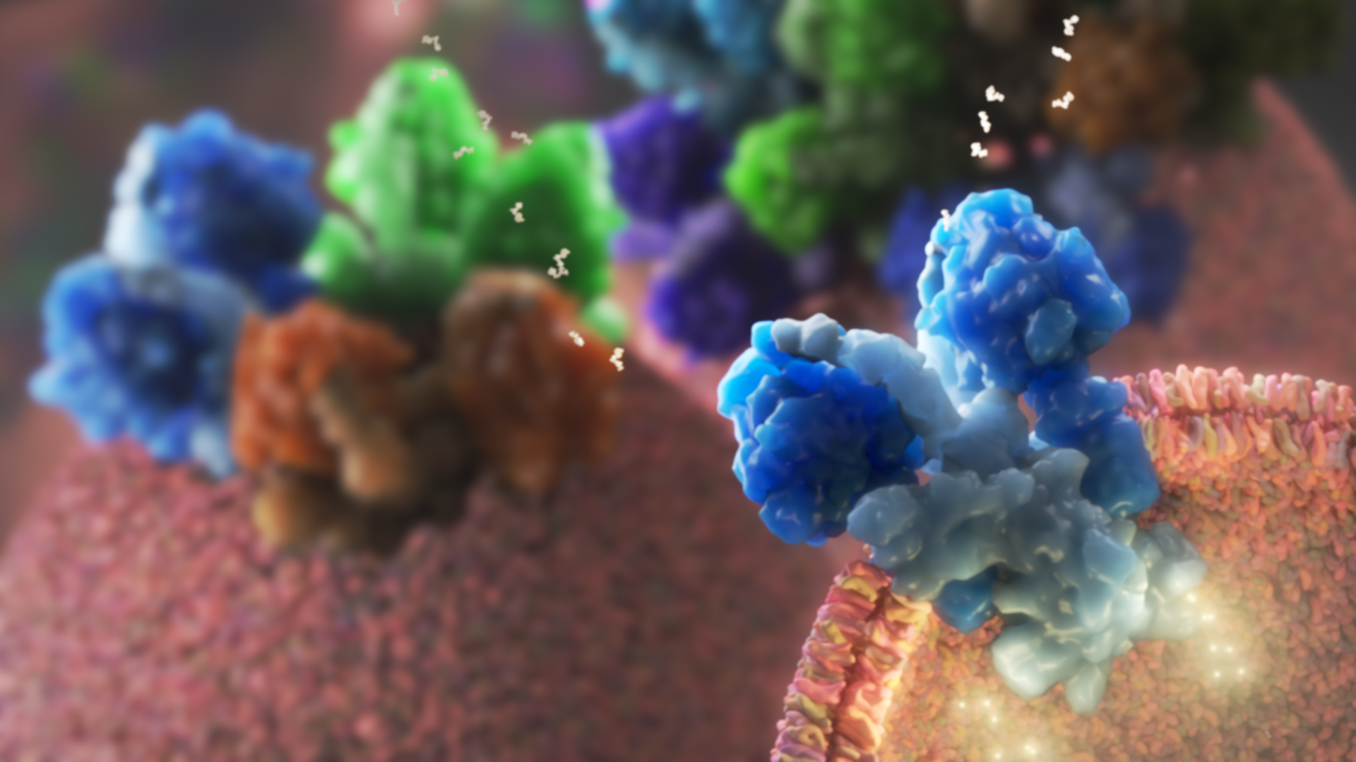
The mitochondrial ATP-synthase is an energy-converting macromolecular machinery that uses the electrochemical potential across the bioenergetic membrane called cristae. This potential is maintained via a membrane curvature that is induced by ATP synthase assembled in dimers. The dimers shaping the bioenergetic membrane were previously thought to be universal across the eukaryotic organisms but two newly published cryo-EM studies by Kock Flygaard et al and Mühleip et al from SciLifeLab Fellow Alexey Amunts lab, have identified different types of ATP synthase organization.
The structure of the ATP synthase from ciliates revealed a dimer which, unlike all the previously investigated complexes, have two membrane-embedded parts that are not identical to each other. The commonly observed symmetry is broken by the accommodation of a single subunit at the dimer interface that anchors an inhibitor. In addition, the ATP synthase has an unusual U-shape arrangement, and thus the generation of the membrane curvature is achieved through tetramerization. This defines the ATP synthase tetramer as the intact structural unit propagating cristae formation in ciliates.
The investigation of the infectious apicomplexan parasites Toxoplasma, revealed that their ATP synthase is arranged in cyclic hexamers. However, within the hexamer, the lipid bilayer turns out to be near-planar, which is not sufficient to shape the bioenergetic membrane. Therefore, the cryo-electron tomography approach was applied to the native membranes isolated from the parasites’ mitochondria, which revealed that the hexamers are further arranged in a higher order of organization. Particularly, 20 units of ATP synthase are linked together in large arrays with icosahedral symmetry.
They form pentagonal pyramids at the size of 20 mega-Dalton. In the center of each pyramid, hexamer ATP synthase planes are oriented by 40°, revealing that the mechanism of pentagonal pyramids generates cristae morphology in a way that differs from the canonical dimers previously thought to be universal.
Finally, the structural studies identified a key subunit, ATPTG11, holding the hexamers together. A removal of the subunit showed loss of pentagonal pyramids, aberrantly shaped cristae, and defective growth of the parasites. This demonstrates that the unique macromolecular arrangement is critical for the maintenance of bioenergetics in Apicomplexa.
Together, these studies illustrate the structural basis for the diversity of the membrane-shaping properties of mitochondrial ATP synthases. This suggests that the fundamental mechanism of the ATP synthase association varies between eukaryotic lineages.
Publications in Nature Communications:
Type III ATP synthase is a symmetry-deviated dimer that induces membrane curvature through tetramerization
ATP synthase hexamer assemblies shape cristae of Toxoplasma mitochondria