
Combining 3D imaging and spatial biology to understand pulmonary hypertension
Pulmonary hypertension is a life-threatening condition for which current treatments can slow disease progression and relieve symptoms, but not target the underlying cause. A demonstrator project within SciLifeLab’s Translation to Healthcare initiative brings together advanced 3D imaging and spatial transcriptomics to understand how the disease develops and pave the way for more precise treatments.
Pulmonary hypertension is characterized by abnormally high pressure in the blood vessels of the lungs. The disease can have several different causes and forms, including pulmonary veno-occlusive disease (PVOD), a rare, severe form affecting children and young adults.
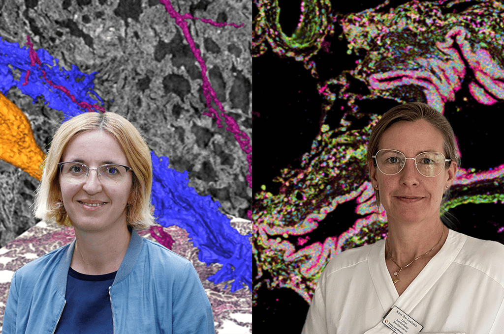
“Most of the treatments available today are vasodilators. They relieve symptoms and improve prognosis but are far from curative. We need more knowledge about the underlying disease mechanisms in different types of pulmonary hypertension,” says Karin Tran-Lundmark, cardiologist at the Pediatric Heart Center at Skåne University Hospital and researcher at Lund University.
One challenge is the complexity of the lung’s vascular network. Conventional tissue analysis provides detailed information from thin, two-dimensional sections that cannot capture how disease related changes are affecting the tissue throughout the blood vessels.
The project combines synchrotron-based phase-contrast micro-CT conducted at the Swedish X-ray facility MAX IV and other similar facilities in Europe, with molecular characterization of the tissue. Imaging in 3D makes it possible to visualize structural changes in lung vessels, and spatial transcriptomics analyses at SciLifeLab adds information about which genes are active, in which cells, and where those cells are located.
“The rationale for using spatial transcriptomics is to gain a better understanding of the molecular and cellular mechanisms underlying pulmonary hypertension,” says Katarina Tiklova, Head of the In Situ Sequencing Unit at SciLifeLab and continues: “By profiling thousands of genes while preserving their spatial location, we can identify the cell types involved and characterize their disease-associated states within the lung tissue. This combination of transcriptomic and spatial information is particularly valuable for understanding the disease in its tissue context.”
The goal is to move from treatments that primarily manage the consequences of disease towards therapies informed by the mechanisms driving disease in the individual patient.
The demonstrator projects developed within Breakthrough Technologies for Health are part of SciLifeLab’s strategic area Translation to Healthcare. The projects explore how emerging molecular technologies, advanced imaging and data analysis can be brought together around concrete clinical challenges, and serve as examples for how research infrastructure and healthcare can work closely together for the benefit of patients.