Industry case: SciLifeLab and AstraZeneca advance collaboration exploring use of NanoSIMS in drug development
A dynamic collaboration between SciLifeLab and AstraZeneca is playing a key role in developing and refining correlative methods as a valuable tool for oligonucleotide drug discovery.
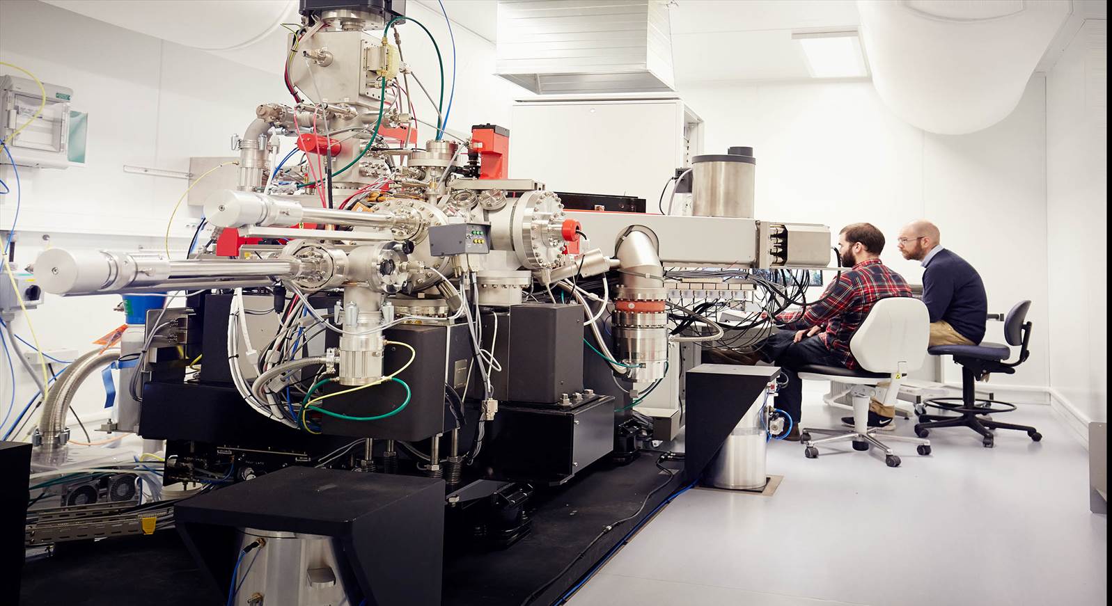
The pharmaceutical industry continues to explore the potential of advanced drug modalities that require intracellular delivery. Understanding how these new therapeutic molecules interact at the subcellular level is critical for drug development. To address this challenge, AstraZeneca and SciLifeLab are using cutting-edge correlative multimodal imaging technology at the SciLifeLab Integrated Microscopy Technology unit in Gothenburg (Centre for Cellular Imaging, University of Gothenburg).
Historically, drug development has largely focused on designing small molecules that circulated in the bloodstream to target specific proteins. Today, the approach has evolved, with, researchers exploring ways to eliminate or modify disease-associated targets within cells. These new approaches often require larger molecules such as oligonucleotides to cross cell membranes and this presents a significant challenge. Imaging technologies such as Electron microscopy and NanoSIMS (nanoscale secondary ion mass spectrometry) are allowing scientists to understand subcellular distribution of these novel investigational therapies by providing high-resolution maps at nanometer scale.
“One of the biggest challenges in drug development is understanding how molecules, in this case oligonucleotides, cross the cell membrane and reach their targets. By leveraging the advanced instrumentation at SciLifeLab, we have developed workflows that allow us to visualize the uptake, distribution and amount of therapeutic molecules with exceptional detail”, says Michael Kurczy, Associate Principal Scientist, AstraZeneca and Adjunct Lecturer, Gothenburg University.
The SciLifeLab Integrated Microscopy Technology unit in Gothenburg, in collaboration with AstraZeneca and the NanoSIMS Sweden facility, has successfully developed, validated, and implemented a correlated multimodal imaging method. This approach combines Electron microscopy and NanoSIMS to precisely track the location of an isotopically labelled, organelle-associated prodrug while directly measuring its absolute concentration. This collaboration has resulted in an innovative quantitative imaging method, specifically designed for the pharmaceutical industry, to offer better understanding the subcellular behaviour of new drug modalities. By combining these technologies with tailored sample preparation, we have obtained striking high-resolution images that enable precise quantification of antisense oligonucleotide levels within individual endosomal structures, says Michael Kurczy*.
As the demand for advanced drug delivery solutions increases, insights gained through this collaboration will contribute to the future of pharmaceutical research. Furthermore, other scientists interested in oligonucleotide therapeutics, intracellular drug delivery, and biomolecular imaging now have a powerful toolset at their disposal to accelerate the development of novel precision medicine approaches.
“The unique strength of this project lies in its broad multidisciplinary approach, bringing together experts from diverse scientific fields. This collaboration is now demonstrating real impact, driving innovation and introducing fresh perspectives to the development of oligonucleotide therapeutics. Companies come to us with specific questions, and we assist them in sample preparation using specialized protocols tailored to various visualization techniques. This allows us to ‘open up the black box’—the cells—providing the pharmaceutical industry with critical insights into how novel therapeutics behave at the subcellular level,” says Julia Fernandez-Rodriguez, SciLifeLab Head of Unit, Integrated Microscopy Technology, University of Gothenburg.
This dynamic collaboration between SciLifeLab and AstraZeneca is playing a key role in developing and refining correlative methods as a valuable tool for oligonucleotide drug discovery. By connecting scientists and expert technical staff from academia and industry across diverse fields, AstraZeneca has introduced a fresh model for collaboration—one that has proven highly effective in driving methodological innovation. The workflows established through this joint effort are now widely available and are being applied to an increasingly broad range of research questions. “This represents a win-win-win scenario: AstraZeneca gains insights that support drug development, SciLifeLab strengthens its capabilities in advanced imaging, and researchers across Sweden benefit from validated methodologies that can be applied to a wide range of biological inquiries,” concludes Julia Fernandez-Rodriguez.
*) Kay E., et al. 2022, Pharmaceutics,14, 463.
Want to know more?

Julia Fernandez-Rodriguez
Head of unit: Integrated Microscopy Technologies, University of Gothenburg

Michael Kurczy
Astra Zeneca
Associate Principal Scientist, AstraZeneca and Adjunct Lecturer, Gothenburg University