Infrastructure update: The Cell and Molecular Imaging platform (CMI)
A new Cell and Molecular Imaging platform (CMI) has been launched, with geographical and technical broadening plans.
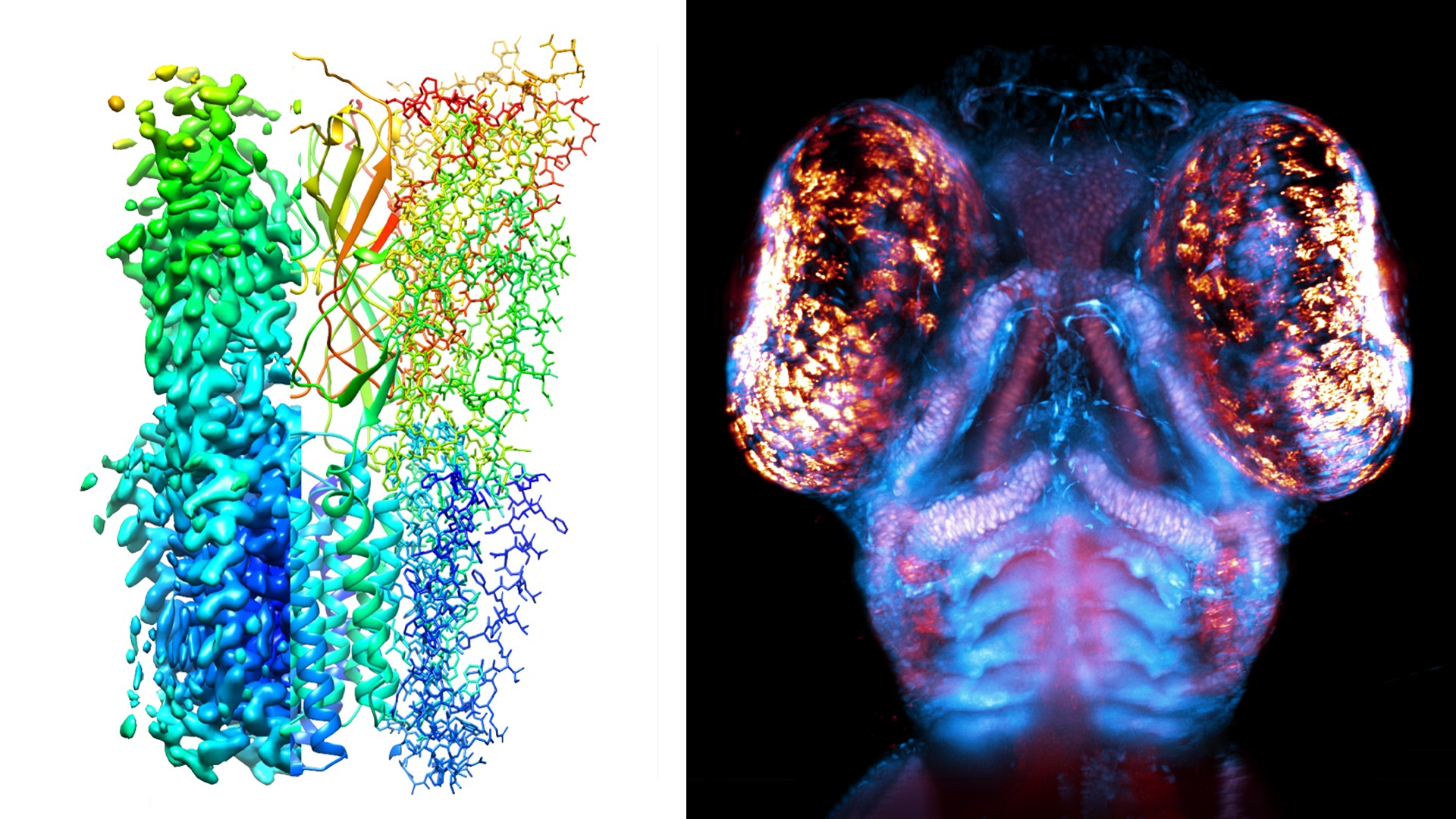
The CMI platform makes the visualization of biological systems, from the molecular to the tissue level possible. Cryo-EM enables the reconstruction of near-atomic structures of molecular machines, super-resolution microscopy makes it possible to produce images with light at the cellular nanoscale, and with expansion and light-sheet microscopy it is possible to reconstruct small organisms or even entire organs, such as kidneys, in detail. The platform is organized in two Units: Cryo-EM and the new IMT (Integrated Microscopy Technologies), each with clear goals for the next four years.
The Cryo-EM Unit, with nodes in Stockholm and Umeå, is developing a national Cryo-EM network, including four new host universities (Uppsala, Lund, Gothenburg and Karolinska Institutet). Staff employed at the host sites, with financial support from SciLifeLab, will be trained by the Cryo-EM Unit and will work as local technical support for starting new projects, thus spreading the technique in Sweden and hopefully “infecting” many new research groups.
The extended IMT Unit (formally known as ALM), in addition to the super-resolution and light-sheet microscopy technologies already on-site, will be hosting two new cutting-edge techniques: FIB-SEM (focused ion beam-scanning electron microscopy) and CAT (correlative array tomography), located in Umeå and Gothenburg, respectively. Bridging electron and photon microscopy, FIB-SEM and CAT will allow the visualization of bigger cellular structures and even larger multicellular 3D volumes. This will illuminate further the network of interaction within and between cells.
With these programs, the CMI hopes to reach out to many new laboratories around Sweden, and possibly, the international arena. One of the researchers that has utilized the platform is Annika Scheynius, professor at Karolinska Institutet. Using imaging tools at the CMI platform, she carried out a study on host-microbe interactions between human skin cells and extracellular nanovesicles released from Malassezia sympodialis, the dominant commensal fungi on human skin.
“It was an excellent collaboration with the cryo-EM and ALM facilities in Stockholm resulting in novel findings”, says Annika.
Photo: Ion channel, Urska Rovsnik (SU) | Zebrafish head, Steven Edwards (KTH)
List of CMI-related publications