
Single-cell imaging in 3D reveals drug differences that flat cultures miss
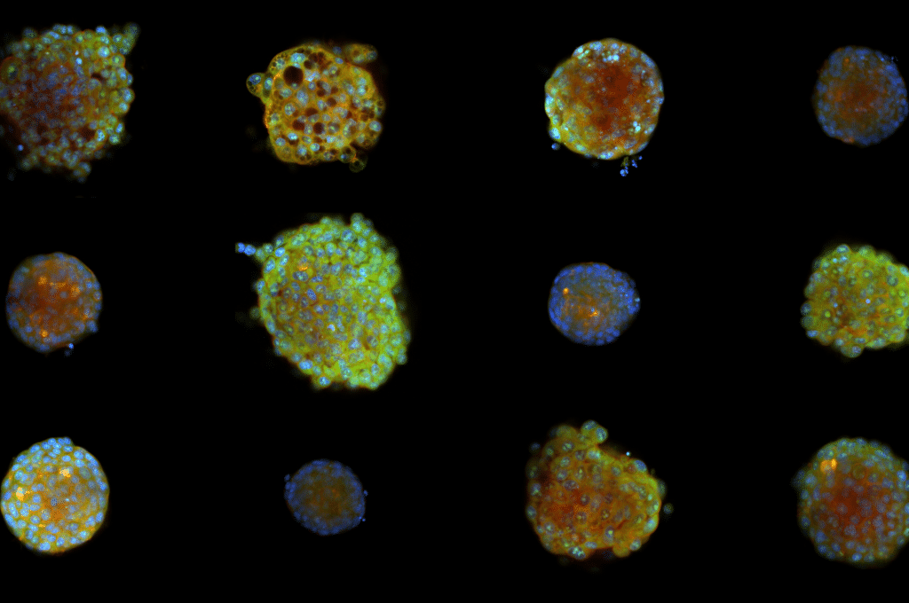
Two cancer drugs that looked almost identical in flat cell cultures turned out to act differently in three-dimensional cell models, but only when you look at individual cells. Christa Ringers and her colleagues found this after adapting Cell Painting, an imaging method used in drug discovery, to study individual cells inside 3D cell spheroids. The study is published in Nature Communications.
Most screening of drug effects is done on cells grown in a single flat layer, even though cells are known to behave differently in three dimensions. Applying a sensitive imaging method like Cell Painting to 3D models at the single-cell level has long been considered difficult.
“Most scalable methods for looking at drug responses run in 2D monolayers, even though people know cells should behave differently in 3D. Getting Cell Painting to work in 3D at single-cell level shouldn’t be impossible when you consider how light moves through samples” says Christa Ringers, senior research engineer at the Department of Pharmaceutical Biosciences, Uppsala University and member of SciLifeLab Group Leader Ola Spjuth’s group.
Hidden in the whole, measured cell by cell
The team made the spheroids transparent to image individual cells throughout the structures. They found that the effects of 5-fluorouracil and olaparib differed in 3D, even though they looked almost identical in conventional 2D cultures and linked the differences to how the drugs activate the cells’ p53 stress response.
Crucially, the distinction was only visible when the researchers looked at individual cells. When they analyzed each spheroid as a single image, the differences disappeared. This suggests that both conventional 2D screens and coarser 3D analyses can miss meaningful differences in how drugs act.
The method runs on equipment that many labs already have, so groups using Cell Painting can add a 3D view with limited new investment. It also works outside the team’s own lab: the researchers ran it at AstraZeneca, which Ringers describes as a particularly rewarding part of the project.
From zebrafish to spheroids
The way the method automatically locates each spheroid in the 3D images borrows an approach used in the zebrafish lab where Ringers previously worked.
“In my earlier zebrafish work, we timed when an odor arrived by finding the moment a fluorescent signal rose fastest. I simply swapped time for depth and brightness for sharpness,” Ringers says.
This study also challenged their expectations about cell growth inside spheroids. A familiar picture of spheroids is an outer layer of growing cells around a core where cells no longer divide. In spheroids as small as the ones in this study, that picture did not hold: cells were dividing all the way to the center.
Collaboration with SciLifeLab
The work was carried out in collaboration with the Chemical Biology Consortium Sweden (CBCS) at SciLifeLab in Uppsala. Experts in 3D cell culture and Cell Painting from the SciLifeLab community also contributed to the study.
Looking ahead, they want to apply the method to more complex 3D models, including organoids grown from patient tissue, and test more cell lines. They also plan to move from analyzing cells slice by slice to measuring them in full 3D.All of the images and data from the study are openly available, and Ringers hope that other researchers will use them to train machine-learning models that can learn drug effects directly from 3D images. They also want to share the method and build expertise in 3D morphological profiling at SciLifeLab.