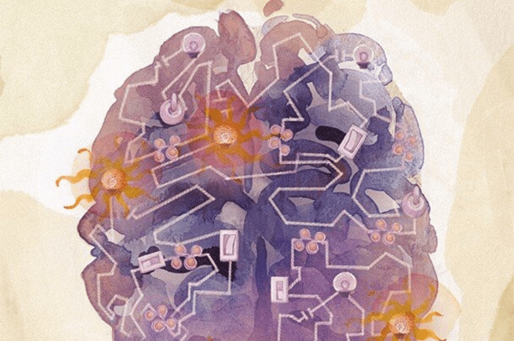
New atlas shows how adult brain cells retain traces of their development
A new study from Karolinska Institutet and SciLifeLab presents a detailed map of how gene activity is regulated in individual cells of the adult human brain and spinal cord. Published in Nature Neuroscience, the study shows that oligodendroglia, the cells responsible for forming myelin, retain an epigenetic “memory” of developmental programs long after these genes are no longer active.
Inside each cell, DNA is tightly packaged together with histone proteins. Gene activity depends on how accessible this packaging is, as well as on chemical modifications to histones that can promote or suppress gene expression.
In this study, researchers used advanced epigenomic methods, including single-nucleus ATAC sequencing and a dual histone profiling technique known as nanoCUT&Tag, to analyze hundreds of thousands of individual cells across multiple regions of the central nervous system.
“For this single-cell epigenomic mapping of the human brain, extensive DNA sequencing was required, which was only possible with the support of the National Genomics Infrastructure at SciLifeLab in Stockholm”, says corresponding author Goncalo Castelo-Branco, Group Leader at SciLifeLab and Professor at KI.
“Having both chromatin accessibility and histone-level information at single cell resolution helps understanding the logic of gene activation and repression in the adult human brain,” he adds.
Previously unknown regulatory region identified
The dataset enabled the researchers to identify a previously unknown DNA region involved in regulating SOX10, a key gene that defines the identity of oligodendrocytes.
The study also shows that adult oligodendrocytes retain epigenetic marks at HOX gene clusters, which play a central role during early development, even though these genes are not active in mature cells.
“This “epigenetic memory” of developmental states may allow adult oligodendrocytes to rapidly activate developmental genes when needed, for example during regeneration, but it may also make them more vulnerable to malignant transformation”, says Dr. Mukund Kabbe, first author of the study.
All datasets are being made publicly available through the UCSC Cell Browser, providing a resource for studying the regulatory landscape of human neural cells.
The research was supported by Cancerfonden, Hjärnfonden, the Knut and Alice Wallenberg Foundation, the Göran Gustafsson Foundation, the Swedish Society for Medical Research, the Swedish Research Council, the European Union, the Chan Zuckerberg Initiative, and the Strategic Research Area Stem Cells and Regenerative Medicine at Karolinska Institutet, among others.
DOI: 10.1038/s41593-026-02208-0
Image: Castelo-Branco lab, Karolinska Institutet, Stockholm, Sweden, Illustration by Amagoia Agirre
Text based on original from Sara Lidman (KI)