Spatial Multimodal Analysis: Combining Mass Spectrometry and next-generation sequencing for advanced brain studies
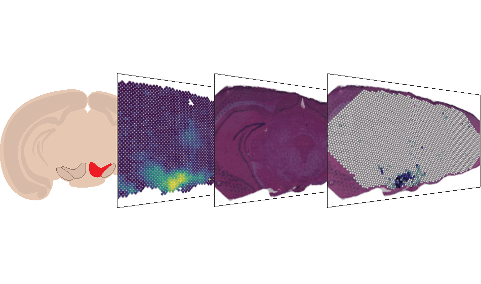
A team of SciLifeLab researchers has introduced a new method, published in Nature Biotechnology, that combines multiple techniques to study a single tissue sample in detail. This combined analysis blends traditional tissue study, imaging techniques that use mass spectrometry, and methods to pinpoint where specific molecules are found in the tissue. By using this approach on mouse and human brain samples, the study explores topics related to neurodegenerative diseases.
”A significant impact of this study is to demonstrate the increased power of spatial multimodality to study events and diseases with subtle molecular differences that occur over extended periods in, for example, neurodegenerative disease,” says Molecular Biotechnology professor Joakim Lundeberg.
This research focuses on two main techniques, supplied by the Spatial Omics service at SciLifeLab, used to study tissues:
- Spatially Resolved Transcriptomics (SRT): This technique looks at the expression of genes throughout a tissue and shows exactly where these genes are active. It’s like getting a map of gene activity across a piece of tissue.
- Mass Spectrometry Imaging (MSI): This method allows researchers to see and measure different molecules in a tissue without labeling them first. A specific type of MSI, called MALDI-MSI, involves coating the tissue with a special substance and then using a laser to detect the molecules present.
“At the best of our knowledge, we have combined the two technologies with the highest throughput in life sciences for the first time: mass spectrometry and next-generation sequencing,” says SciLifeLab researcher and KTH PhD student Marco Vicari.
Although both techniques offer unique insights, they are usually performed separately. This is because of technical challenges and risks involved, like potential damage to the tissue’s RNA – a vital molecule for studying gene activity.
However, in this research, a new method has been introduced that combines both SRT and MALDI-MSI. This method, Spatial Multimodal Analysis (SMA), allows researchers to study tissues with both techniques simultaneously without compromising the quality or accuracy of the results.
”Our method enables imaging and increased precision in measurements of low-molecular-weight metabolites and RNA transcripts in the same biological tissue. The study’s findings have the potential to significantly improve the field of spatial biology and disease research,” says Per Andrén, professor of Imaging Mass Spectrometry at Uppsala University and SciLifeLab.
DOI: s41587-023-01937-y
