

Briefly about Cellular and Molecular Imaging
Cellular and molecular imaging enables the visualization of biological systems from atomistic to tissue levels. Typical methods involve the transmission, absorption, reflection, or refraction of a focused beam of visible light or electrons by a microscopic sample of interest. In the life sciences, such samples may be biological macromolecules, cells, small organs or organisms of interest to biomedical or planetary biology research. At SciLifeLab, imaging staff assist users from academic, industry, and clinical settings to identify and implement relevant techniques for sample preparation and for the collection, processing, analysis, and sharing of imaging data, with a focus on methods and instruments not generally accessible to individual research groups or local facilities. Key SciLifeLab imaging services include cryogenic electronic microscopy (cryo-EM) and tomography (cryo-ET), super-resolution and other advanced methods in fluorescence microscopy, focused ion-beam milling scanning electron microscopy (FIB-SEM), correlative array tomography (CAT) and nanoscale secondary ion mass spectrometry (nanoSIMS), along with a range of imaging modalities in support of structural biology and spatial omics.
Contact person
For general inquiries about cellular and molecular imaging services at SciLifeLab, please contact Reba.

Platform Coordination Officer
Cellular and Molecular Imaging Platform
KTH Royal Institute of Technology
cmi-service@scilifelab.se
+46 (0) 72 007 94 51
What we offer at SciLifeLab
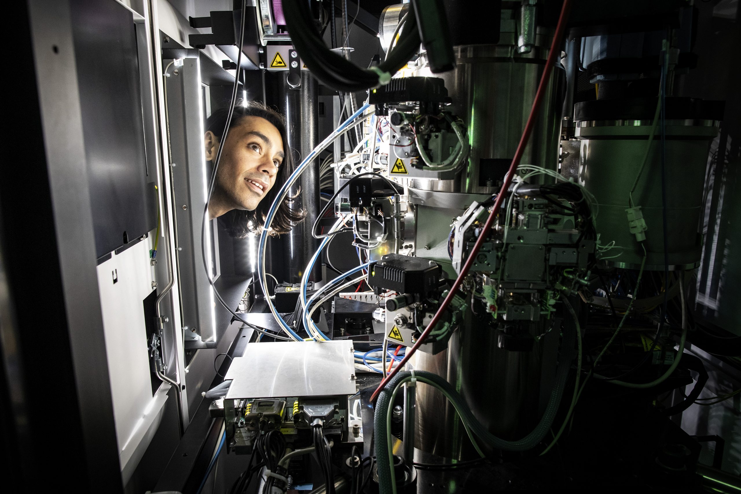
Cryogenic Electron Microscopy Imaging
Cryo-EM is a widely used method for imaging samples embedded and flash-frozen in vitrified ice. Single-particle analysis (SPA) enables structure determination of macromolecules to near-atomic resolution, while cryogenic electron tomography (cryo-ET) reveals regional volumes containing molecules, subcellular structures, or entire cells. With microscopes in Stockholm and Umeå, a network of sample preparation support in Gothenburg, Lund, and Uppsala, and dedicated data-processing specialists, the Cryo-EM unit offers imaging services and support in:
- Cryo-EM grid preparation and data collection for SPA
- Cryo-ET sample preparation and data collection
- Cryogenic correlative light and electron microscopy (cryo-CLEM)
- Microcrystal electron diffraction (micro-ED)
- Data processing and molecular modeling
Explore more
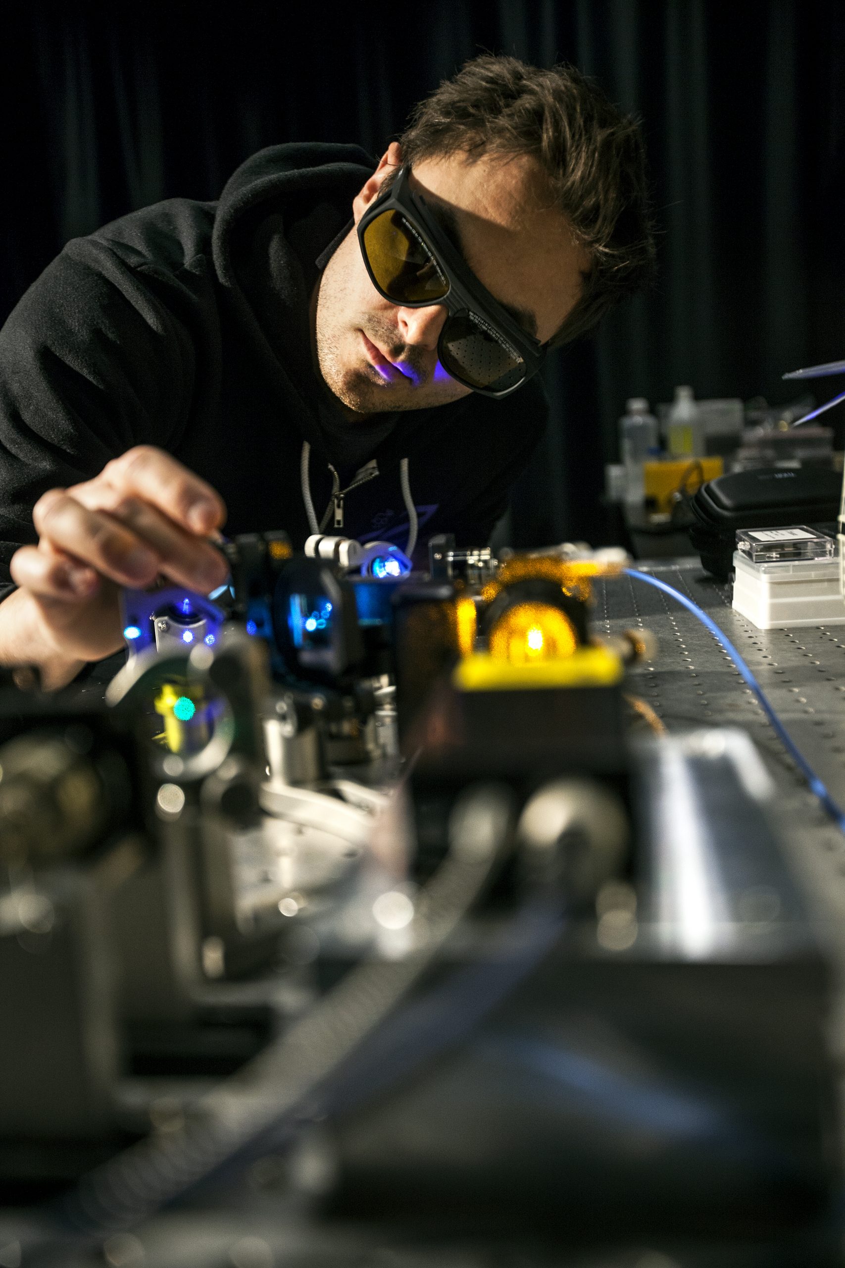
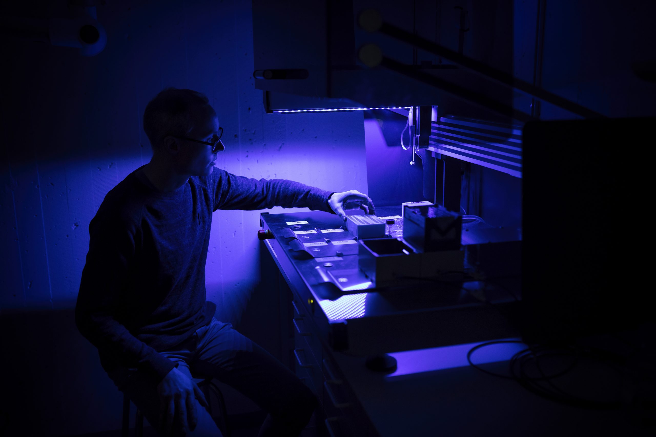
Advanced Light Microscopy Imaging
Advanced fluorescence methods make it possible to visualize nanoscale biostructures, track single-molecule dynamics for example by fluorescence correlation spectroscopy (FCS), and process ultrafast high-resolution 3D images. The Stockholm node of the Integrated Microscopy Technologies unit offers imaging support in:
- Fast volumetric light microscopy and clearing
- Light-sheet and expansion microscopy
- Single-molecule dynamic measurements (e.g. FCS)
- Super-resolution fluorescence microscopy imaging
(e.g. STED, PALM, STORM, SIMS, MINFLUX)
Explore more
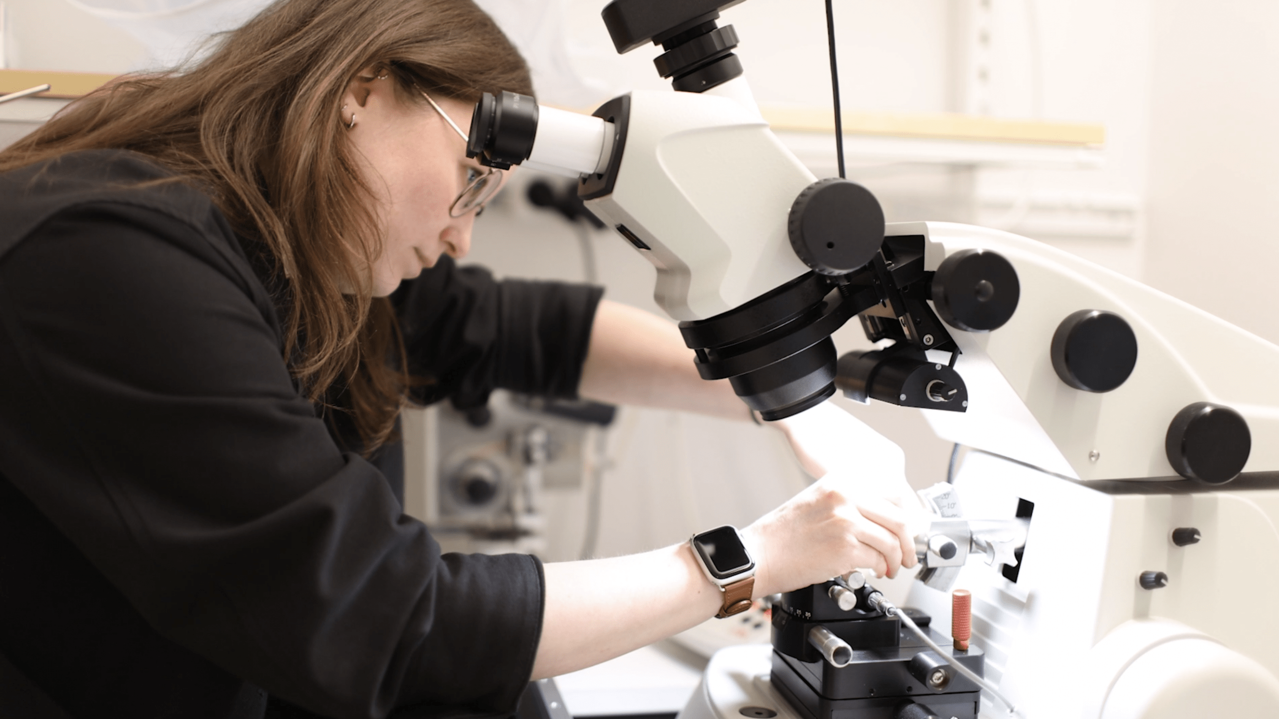
Room-Temperature Light/Electron Microscopy Imaging
Integrating electron microscopy methods with fluorescence imaging or ion-beam milling can reveal details of tissue or cellular structures by leveraging the strengths of both modalities. The Umeå and Gothenburg nodes of the Integrated Microscopy Technologies unit offer imaging support in:
- Correlative array tomography (CAT)
- Correlative light and electron microscopy (CLEM)
- Focused ion beam scanning electron microscopy (FIB-SEM)
Explore more
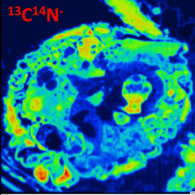
Mass Spectrometry Imaging
Nanoscale secondary ion mass spectrometry (NanoSIMS) measures elemental, isotopic, inorganic, organic, and biomolecular distributions in a broad range of samples with ppm sensitivity, and 2D/3D imaging with nanometer spatial resolution. The Gothenburg node of the Integrated Microscopy Technologies unit offers imaging support in:
- Sample preparation and routine measurements with nanoSIMS
- Training for data processing and analysis of 2D and 3D images
Explore more
Structure Imaging by NMR
Like other core methods of structural biology, nuclear magnetic resonance (NMR) spectroscopy is a powerful tool to determine the physical shape and interactions of biological macromolecules—in effect, imaging on the molecular to atomistic scales. NMR is particularly applicable to soluble proteins and nucleic acids of relatively small size (≤25 kilodaltons), typically in the context of isotopic labeling. With nodes in Gothenburg and Umeå, the Swedish NMR Centre provides service and support for structural biology and metabolomics studies in liquid and solid phase, as well as fragment-based screening.
- Macromolecular structure determination by NMR
- Metabolomics
- Chemical biology and small-molecule NMR
- Solid state/dynamic nuclear polarization (DNP) NMR
Explore more
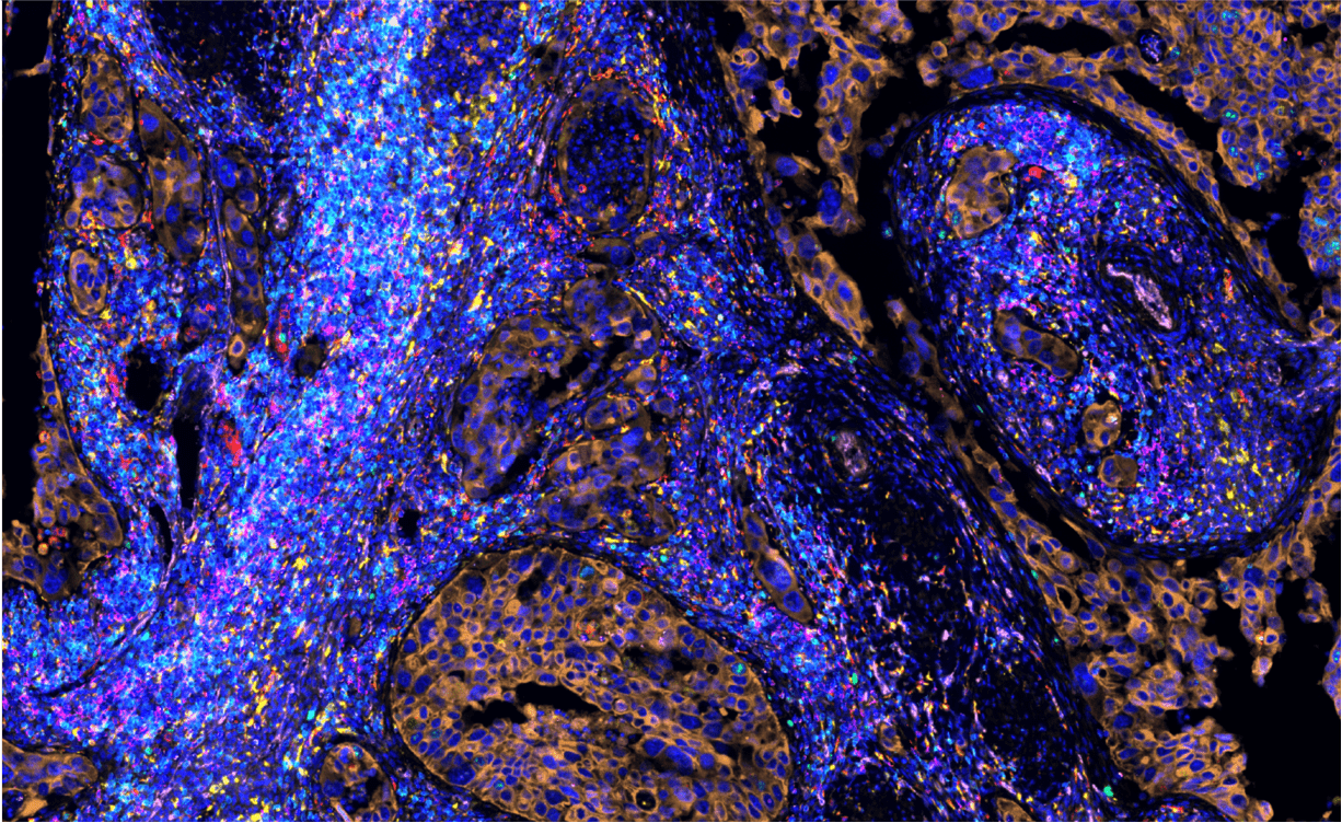
Imaging in Spatial Omics
Spatially resolved omics technologies have provided new understanding of the spatial organization of complex multicellular biological systems. Given its focus on bioprocesses differentiated in space, many spatial omics approaches fundamentally involve the integration of imaging data with other modalities. At SciLifeLab, the Spatial and Single Cell Biology platform provides access and support in cutting-edge technologies for spatial profiling of transcripts, proteins, DNA, and small molecules, as well as several approaches for single-cell analysis.
- Spatial proteomics
- Spatial transcriptomics
- Spatial mass spectrometry
- Advanced fluorescence in-situ hybridization technologies
- In-situ sequencing
Explore more
Bioinformatics Support
The SciLifeLab Bioinformatics platform (NBIS) offers support to imaging projects, such as:
- Free drop-in and initial project consultation
- Embedded support for structural biology data processing
- Bioimage analysis using computer vision, machine learning, and bioinformatics
- Protein structure prediction including AlphaFold2
- Open Science, FAIR data management, and data publication support
Explore more
Other Infrastructure Resources
Access to equipment and competence in microscopy for the life science research community has been provided since 2016 by the Swedish National Microscopy Infrastructure (NMI). With sites in Stockholm, Umeå, Gothenburg, and Uppsala, NMI coordinates national and international knowledge exchange in microscopy, and is the Swedish node of EuroBioimaging.
- Advanced methods including super-resolution, single-molecule and light-sheet
- Live and intravital imaging of cells, tissues, organs, biomaterials
- Multimodal and correlative approaches
Synchrotron radiation produces X-rays with high brilliance, polarization, and tunability, enabling distinctive applications in bioimaging. The Swedish national synchrotron MAX IV Laboratory in Lund has welcomed users since 2016 to 16 beamlines, 4 of which (SoftiMAX, MedMAX, NanoMAX, ForMAX) specialize in bioimaging among other applications.
Neutrons constitute unique probes for investigating the structure and function of matter, from microscopic to atomic scales. Facilitated by the brightness of the newly launched European Spallation Source (ESS) in Lund, and empowered by ongoing advances in detector technology. the optical diffraction imaging with neutrons (ODIN) instrument enables imaging down to the µm range.
- Traditional attenuation-based to advanced dark-field imaging
- Wavelength resolution, bandwidth and collimation tailored to each application