Heart ‘blueprint’ made openly available in unique resource
A detailed map of the developing human heart, showing how different groups of cells are arranged and how they interact in fetal heart development have been published by a team led by SciLifeLab and KTH Royal Institute of Technology researchers.
“Congenital heart diseases, and several acquired ones, originate during early development, which highlights the importance of this period in defining a healthy heart,” says Enikő Lázár, KTH and SciLifeLab researcher and co-lead author of the study, in a KTH press release, “This map provides a kind of blueprint, showing how key parts of the heart—like the pacemaker system, heart valves, and the wall between the upper chambers—form and function”.
A previously unknown group of cells likely unique to humans that produce adrenaline was also found. These cells may help the heart respond to low oxygen levels during development or birth.
“Our collaborative study offers a unique resource to explore the cellular and molecular blueprint of human heart development, offering new links to genetic causes of heart disease. Importantly, our spatial results are available through an open-access spatially centric interactive viewer developed and hosted by SciLifeLab (TissUUmaps) for further explorations by the scientific community,” says SciLifeLab Group Leader and KTH researcher Joakim Lundeberg.
Spatial transcriptomics enabled visualization and analysis of the full range of messenger RNA, or mRNA.
“With careful bioinformatic analysis, we can learn details of heart development that were not possible even a few years ago,” says Raphaël Mauron, KTH and SciLifeLab researcher and co-lead author of the study, in a KTH press release.
DOI: 10.1038/s41588-025-02352-6
Learn more in the KTH press release
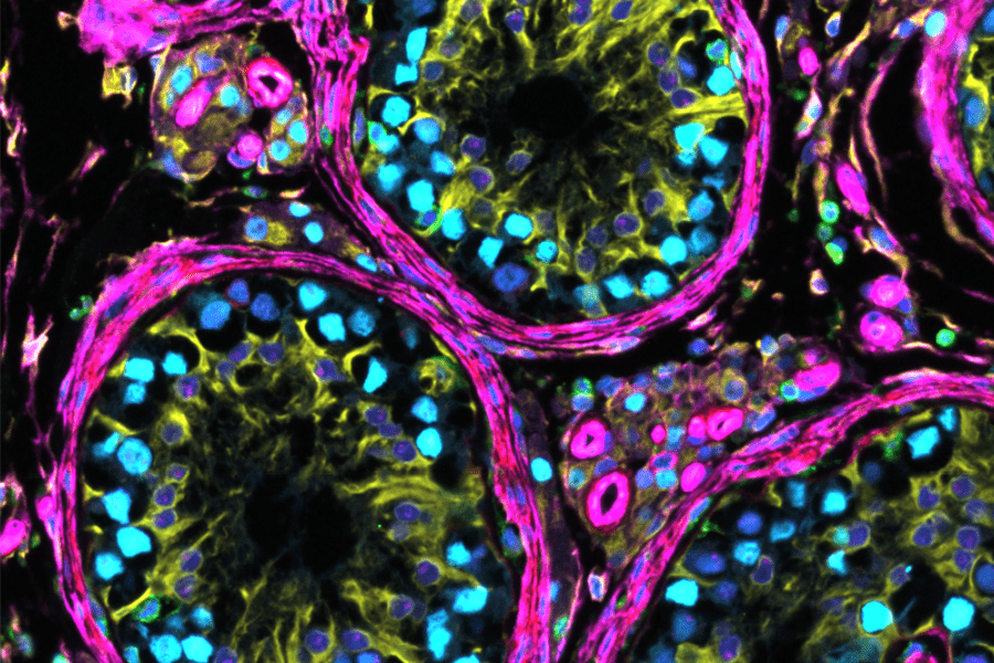
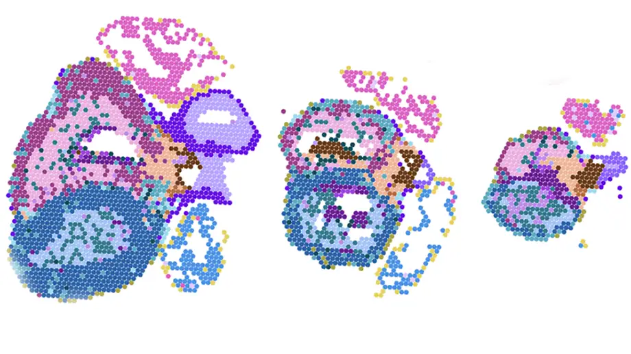
Image: A visualization of molecular data generated from three distinct points in time during fetal heart formation.
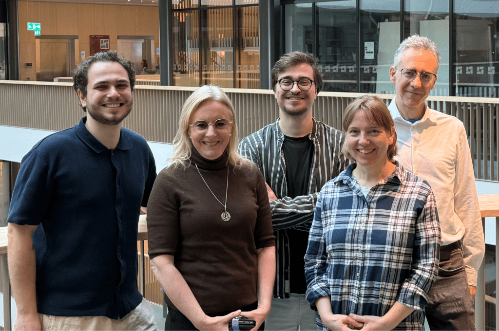
Photo of Enikő Lázár & Raphaël Mauron: Patrick Truong.